[English] 日本語
Yorodumi
- PDB-9arn: Structure and interactions of HIV-1 gp41 CHR-NHR reverse hairpin ... -

+
Open data


ID or keywords:

Loading...

-
Basic information

Entry
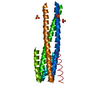
Database: PDB / ID: 9arn
TitleStructure and interactions of HIV-1 gp41 CHR-NHR reverse hairpin constructs reveal molecular determinants of antiviral activity
ComponentsTransmembrane protein gp41
KeywordsVIRAL PROTEIN / HIV / GP41 / Virus / Reverse Hairpin
Function / homology
Function and homology information


Synthesis and processing of ENV and VPU / symbiont-mediated evasion of host immune response / Alpha-defensins / Dectin-2 family / Binding and entry of HIV virion / positive regulation of plasma membrane raft polarization / positive regulation of receptor clustering / positive regulation of establishment of T cell polarity / host cell endosome membrane / actin filament organization ...Synthesis and processing of ENV and VPU / symbiont-mediated evasion of host immune response / Alpha-defensins / Dectin-2 family / Binding and entry of HIV virion / positive regulation of plasma membrane raft polarization / positive regulation of receptor clustering / positive regulation of establishment of T cell polarity / host cell endosome membrane / actin filament organization / Assembly Of The HIV Virion / Budding and maturation of HIV virion / clathrin-dependent endocytosis of virus by host cell / viral protein processing / symbiont entry into host cell / fusion of virus membrane with host plasma membrane / fusion of virus membrane with host endosome membrane / : / viral envelope / virion attachment to host cell / apoptotic process / host cell plasma membrane / virion membrane / structural molecule activity / membrane
Similarity search - Function
Envelope glycoprotein Gp160 / Retroviral envelope protein / Retroviral envelope protein GP41-like / Gp120 core superfamily / Envelope glycoprotein GP120 / Human immunodeficiency virus 1, envelope glycoprotein Gp120
Similarity search - Domain/homology
Envelope glycoprotein gp160
Similarity search - Component
Biological speciesHuman immunodeficiency virus 1
MethodX-RAY DIFFRACTION / SYNCHROTRON / MOLECULAR REPLACEMENT / Resolution: 1.41 Å
AuthorsMcAndrew, R.P. / Ralston, C.Y. / Gochin, M.
Funding support United States, 3items
OrganizationGrant numberCountry
National Institutes of Health/National Institute Of Allergy and Infectious Diseases (NIH/NIAID)R21 AI140904 United States
National Institutes of Health/National Institute of General Medical Sciences (NIH/NIGMS)1R01GM126218 United States
National Institutes of Health/National Institute of General Medical Sciences (NIH/NIGMS)P30 GM124169 United States
Citation
Journal: J.Mol.Biol. / Year: 2024
Title: Structure and Interactions of HIV-1 gp41 CHR-NHR Reverse Hairpin Constructs Reveal Molecular Determinants of Antiviral Activity.
Authors: He, L. / McAndrew, R. / Barbu, R. / Gifford, G. / Halacoglu, C. / Drouin-Allaire, C. / Weber, L. / Kristensen, L.G. / Gupta, S. / Chen, Y. / Petzold, C.J. / Allaire, M. / Li, K.H. / Ralston, C.Y. / Gochin, M.
#1: Journal: Acta Crystallogr D Struct Biol / Year: 2019
Title: Macromolecular structure determination using X-rays, neutrons and electrons: recent developments in Phenix.
Authors: Dorothee Liebschner / Pavel V Afonine / Matthew L Baker / Gábor Bunkóczi / Vincent B Chen / Tristan I Croll / Bradley Hintze / Li Wei Hung / Swati Jain / Airlie J McCoy / Nigel W Moriarty ...Authors: Dorothee Liebschner / Pavel V Afonine / Matthew L Baker / Gábor Bunkóczi / Vincent B Chen / Tristan I Croll / Bradley Hintze / Li Wei Hung / Swati Jain / Airlie J McCoy / Nigel W Moriarty / Robert D Oeffner / Billy K Poon / Michael G Prisant / Randy J Read / Jane S Richardson / David C Richardson / Massimo D Sammito / Oleg V Sobolev / Duncan H Stockwell / Thomas C Terwilliger / Alexandre G Urzhumtsev / Lizbeth L Videau / Christopher J Williams / Paul D Adams /
Abstract: Diffraction (X-ray, neutron and electron) and electron cryo-microscopy are powerful methods to determine three-dimensional macromolecular structures, which are required to understand biological ...Diffraction (X-ray, neutron and electron) and electron cryo-microscopy are powerful methods to determine three-dimensional macromolecular structures, which are required to understand biological processes and to develop new therapeutics against diseases. The overall structure-solution workflow is similar for these techniques, but nuances exist because the properties of the reduced experimental data are different. Software tools for structure determination should therefore be tailored for each method. Phenix is a comprehensive software package for macromolecular structure determination that handles data from any of these techniques. Tasks performed with Phenix include data-quality assessment, map improvement, model building, the validation/rebuilding/refinement cycle and deposition. Each tool caters to the type of experimental data. The design of Phenix emphasizes the automation of procedures, where possible, to minimize repetitive and time-consuming manual tasks, while default parameters are chosen to encourage best practice. A graphical user interface provides access to many command-line features of Phenix and streamlines the transition between programs, project tracking and re-running of previous tasks.
History
DepositionFeb 23, 2024Deposition site: RCSB / Processing site: RCSB
Revision 1.0Mar 5, 2025Provider: repository / Type: Initial release

-
Structure visualization

Structure viewerMolecule:
MolmilJmol/JSmol

Downloads & links

-
Assembly

Deposited unit
A: Transmembrane protein gp41
hetero molecules


Theoretical massNumber of molelcules
Total (without water)9,6522
Polymers9,5561
Non-polymers961
Water68538
1
A: Transmembrane protein gp41
hetero molecules

A: Transmembrane protein gp41
hetero molecules

A: Transmembrane protein gp41
hetero molecules


Theoretical massNumber of molelcules
Total (without water)28,9556
Polymers28,6673
Non-polymers2883
Water543
TypeNameSymmetry operationNumber
identity operation1_555x,y,z1
crystal symmetry operation2_555-y,x-y,z1
crystal symmetry operation3_555-x+y,-x,z1
MethodPISA
Unit cell
Length a, b, c (Å)44.783, 44.783, 220.058
Angle α, β, γ (deg.)90.000, 90.000, 120.000
Int Tables number155
Space group name H-MH32
Space group name HallR32"
Symmetry operation#1: x,y,z
#2: -y,x-y,z
#3: -x+y,-x,z
#4: x-y,-y,-z
#5: -x,-x+y,-z
#6: y,x,-z
#7: x+1/3,y+2/3,z+2/3
#8: -y+1/3,x-y+2/3,z+2/3
#9: -x+y+1/3,-x+2/3,z+2/3
#10: x-y+1/3,-y+2/3,-z+2/3
#11: -x+1/3,-x+y+2/3,-z+2/3
#12: y+1/3,x+2/3,-z+2/3
#13: x+2/3,y+1/3,z+1/3
#14: -y+2/3,x-y+1/3,z+1/3
#15: -x+y+2/3,-x+1/3,z+1/3
#16: x-y+2/3,-y+1/3,-z+1/3
#17: -x+2/3,-x+y+1/3,-z+1/3
#18: y+2/3,x+1/3,-z+1/3
Components on special symmetry positions
IDModelComponents
11A-201-

HOH

21A-226-

HOH

31A-235-

HOH

-
Components

#1: Protein Transmembrane protein gp41 / TM / Glycoprotein 41 / gp41


Mass: 9555.728 Da / Num. of mol.: 1
Fragment: residues 542-591 (Uniprot numbering), plus N-terminal extension
Source method: isolated from a genetically manipulated source
Source: (gene. exp.) Human immunodeficiency virus 1 / Gene: env / Production host: Escherichia coli (E. coli) / References: UniProt: P04578
#2: Chemical ChemComp-SO4 / SULFATE ION


Mass: 96.063 Da / Num. of mol.: 1 / Source method: obtained synthetically / Formula: SO4
#3: Water ChemComp-HOH / water


Mass: 18.015 Da / Num. of mol.: 38 / Source method: isolated from a natural source / Formula: H2O
Has ligand of interestN
Has protein modificationN

-
Experimental details

-
Experiment

ExperimentMethod: X-RAY DIFFRACTION / Number of used crystals: 1

-
Sample preparation

CrystalDensity Matthews: 2.22 Å3/Da / Density % sol: 44.65 %
Crystal growTemperature: 293 K / Method: vapor diffusion / pH: 6.4
Details: 0.1M lithium sulfate, 0.1M sodium citrate trihydrate at pH 6.4 and 25% PEG-1500

-
Data collection

DiffractionMean temperature: 100 K / Serial crystal experiment: N
Diffraction sourceSource: SYNCHROTRON / Site: ALS / Beamline: 5.0.1 / Wavelength: 1.00003 Å
DetectorType: DECTRIS PILATUS3 6M / Detector: PIXEL / Date: Mar 18, 2022
RadiationProtocol: SINGLE WAVELENGTH / Monochromatic (M) / Laue (L): M / Scattering type: x-ray
Radiation wavelengthWavelength: 1.00003 Å / Relative weight: 1
ReflectionResolution: 1.41→38.19 Å / Num. obs: 18490 / % possible obs: 92.13 % / Redundancy: 8.2 % / Biso Wilson estimate: 21.45 Å2 / CC1/2: 1 / CC star: 1 / Rmerge(I) obs: 0.04578 / Net I/σ(I): 8.71
Reflection shellResolution: 1.41→1.5 Å / Redundancy: 6 % / Rmerge(I) obs: 1.368 / Mean I/σ(I) obs: 0.11 / Num. unique obs: 2999 / CC1/2: 0.64 / CC star: 0.883 / % possible all: 53.02

-
Processing

Software
NameVersionClassification
PHENIXdev_5246refinement
xia20.5.653-g9f819c0c-dials-1.11data reduction
xia20.5.653-g9f819c0c-dials-1.11data scaling
PHASER2.8.3phasing
RefinementMethod to determine structure: MOLECULAR REPLACEMENT / Resolution: 1.41→38.19 Å / SU ML: 0.3208 / Cross valid method: FREE R-VALUE / σ(F): 1.33 / Phase error: 46.3373
Stereochemistry target values: GeoStd + Monomer Library + CDL v1.2
RfactorNum. reflection% reflection
Rfree0.2912 771 4.95 %
Rwork0.2555 14805 -
obs0.2573 15576 92.13 %
Solvent computationShrinkage radii: 0.9 Å / VDW probe radii: 1.1 Å / Solvent model: FLAT BULK SOLVENT MODEL
Displacement parametersBiso mean: 39.09 Å2
Refinement stepCycle: LAST / Resolution: 1.41→38.19 Å
ProteinNucleic acidLigandSolventTotal
Num. atoms606 0 5 38 649
Refine LS restraints
Refine-IDTypeDev idealNumber
X-RAY DIFFRACTIONf_bond_d0.0062619
X-RAY DIFFRACTIONf_angle_d0.7259832
X-RAY DIFFRACTIONf_chiral_restr0.045595
X-RAY DIFFRACTIONf_plane_restr0.0071109
X-RAY DIFFRACTIONf_dihedral_angle_d21.2748251
LS refinement shell
Resolution (Å)Rfactor RfreeNum. reflection RfreeRfactor RworkNum. reflection RworkRefine-ID% reflection obs (%)
1.41-1.50.4461770.49351382X-RAY DIFFRACTION53.02
1.5-1.620.42011220.40642635X-RAY DIFFRACTION99.24
1.62-1.780.35641410.34442621X-RAY DIFFRACTION99.78
1.78-2.040.31081480.2752663X-RAY DIFFRACTION99.96
2.04-2.570.24921240.24072690X-RAY DIFFRACTION99.96
2.57-38.190.27911590.22452814X-RAY DIFFRACTION99.8
Refinement TLS params.

Method: refined / Refine-ID: X-RAY DIFFRACTION

IDL112)L122)L132)L222)L232)L332)S11 (Å °)S12 (Å °)S13 (Å °)S21 (Å °)S22 (Å °)S23 (Å °)S31 (Å °)S32 (Å °)S33 (Å °)T112)T122)T132)T222)T232)T332)Origin x (Å)Origin y (Å)Origin z (Å)
14.72734137136-0.3509966633181.394446749523.78743891585-0.268947995913.25156107592-0.0533888667023-0.3310726726130.3565905885460.748818379021-0.191440825392-0.893186432295-0.6064372343381.061327926770.1454183896160.439718404732-0.0378708840804-0.07912245504870.62425062248-0.09534832892190.37978696058811.20455414943.744692729581.8352126561
21.18282790742-0.07045208533141.401078709560.6354058800210.0277684663976.148733184240.0123632699924-0.2970842571480.0594964330690.2700868207310.0513326325774-0.00307468349916-0.4449260017-0.368385581251-0.04070568086090.2514506787550.02248308215150.03501548878410.230618805168-0.009622873069870.180173574852-2.607369424684.3053701391558.127846818
Refinement TLS group

Refine-ID: X-RAY DIFFRACTION / Auth asym-ID: A / Label asym-ID: A

IDRefine TLS-IDSelection detailsAuth seq-IDLabel seq-ID
11chain 'A' and (resid 1 through 31 )1 - 311 - 23
22chain 'A' and (resid 32 through 80 )32 - 8024 - 72

+
About Yorodumi

-
News

-
Feb 9, 2022. New format data for meta-information of EMDB entries

New format data for meta-information of EMDB entries

  • Version 3 of the EMDB header file is now the official format.
  • The previous official version 1.9 will be removed from the archive.

Related info.:EMDB header

External links:wwPDB to switch to version 3 of the EMDB data model

-
Aug 12, 2020. Covid-19 info

Covid-19 info

URL: https://pdbj.org/emnavi/covid19.php

New page: Covid-19 featured information page in EM Navigator.

Related info.:Covid-19 info / Mar 5, 2020. Novel coronavirus structure data

+
Mar 5, 2020. Novel coronavirus structure data

Novel coronavirus structure data

Related info.:Yorodumi Speices / Aug 12, 2020. Covid-19 info

External links:COVID-19 featured content - PDBj / Molecule of the Month (242):Coronavirus Proteases

+
Jan 31, 2019. EMDB accession codes are about to change! (news from PDBe EMDB page)

EMDB accession codes are about to change! (news from PDBe EMDB page)

  • The allocation of 4 digits for EMDB accession codes will soon come to an end. Whilst these codes will remain in use, new EMDB accession codes will include an additional digit and will expand incrementally as the available range of codes is exhausted. The current 4-digit format prefixed with “EMD-” (i.e. EMD-XXXX) will advance to a 5-digit format (i.e. EMD-XXXXX), and so on. It is currently estimated that the 4-digit codes will be depleted around Spring 2019, at which point the 5-digit format will come into force.
  • The EM Navigator/Yorodumi systems omit the EMD- prefix.

Related info.:Q: What is EMD? / ID/Accession-code notation in Yorodumi/EM Navigator

External links:EMDB Accession Codes are Changing Soon! / Contact to PDBj

+
Jul 12, 2017. Major update of PDB

Major update of PDB

  • wwPDB released updated PDB data conforming to the new PDBx/mmCIF dictionary.
  • This is a major update changing the version number from 4 to 5, and with Remediation, in which all the entries are updated.
  • In this update, many items about electron microscopy experimental information are reorganized (e.g. em_software).
  • Now, EM Navigator and Yorodumi are based on the updated data.

External links:wwPDB Remediation / Enriched Model Files Conforming to OneDep Data Standards Now Available in the PDB FTP Archive

-
Yorodumi

Thousand views of thousand structures

  • Yorodumi is a browser for structure data from EMDB, PDB, SASBDB, etc.
  • This page is also the successor to EM Navigator detail page, and also detail information page/front-end page for Omokage search.
  • The word "yorodu" (or yorozu) is an old Japanese word meaning "ten thousand". "mi" (miru) is to see.

Related info.:EMDB / PDB / SASBDB / Comparison of 3 databanks / Yorodumi Search / Aug 31, 2016. New EM Navigator & Yorodumi / Yorodumi Papers / Jmol/JSmol / Function and homology information / Changes in new EM Navigator and Yorodumi

Read more