 Movie
Movie Controller

[English] 日本語
 Yorodumi
- PDB-9aat: X-RAY STRUCTURE REFINEMENT AND COMPARISON OF THREE FORMS OF MITOC... -
+ Open data

- Basic information
| Entry | Database: PDB / ID: 9aat | ||||||
|---|---|---|---|---|---|---|---|
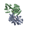
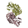
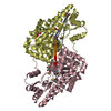
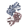
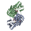
| Title | X-RAY STRUCTURE REFINEMENT AND COMPARISON OF THREE FORMS OF MITOCHONDRIAL ASPARTATE AMINOTRANSFERASE | ||||||
 Components Components | ASPARTATE AMINOTRANSFERASE | ||||||
 Keywords Keywords | TRANSFERASE(AMINOTRANSFERASE) | ||||||
| Function / homology |  Function and homology information Function and homology informationAmino acid metabolism / Gluconeogenesis / L-aspartate catabolic process / kynurenine-oxoglutarate transaminase / L-kynurenine:2-oxoglutarate transaminase activity / aspartate metabolic process / glutamate metabolic process / aspartate transaminase / L-aspartate:2-oxoglutarate transaminase activity / 2-oxoglutarate metabolic process ...Amino acid metabolism / Gluconeogenesis / L-aspartate catabolic process / kynurenine-oxoglutarate transaminase / L-kynurenine:2-oxoglutarate transaminase activity / aspartate metabolic process / glutamate metabolic process / aspartate transaminase / L-aspartate:2-oxoglutarate transaminase activity / 2-oxoglutarate metabolic process / pyridoxal phosphate binding / mitochondrial matrix / protein homodimerization activity / mitochondrion Similarity search - Function | ||||||
| Biological species |  | ||||||
| Method |  X-RAY DIFFRACTION / Resolution: 2.2 Å X-RAY DIFFRACTION / Resolution: 2.2 Å | ||||||
 Authors Authors | Mcphalen, C.A. / Vincent, M.G. / Jansonius, J.N. | ||||||
 Citation Citation | Journal: J.Mol.Biol. / Year: 1992 Title: X-ray structure refinement and comparison of three forms of mitochondrial aspartate aminotransferase. Authors: McPhalen, C.A. / Vincent, M.G. / Jansonius, J.N. #1: Journal: Eur.J.Biochem. / Year: 1991Title: The Open(Slash)Closed Conformational Equilibrium of Aspartate Aminotransferase: Studies in the Crystalline State and with a Fluorescent Probe in Solution Authors: Picot, D. / Sandmeier, E. / Thaller, C. / Vincent, M.G. / Christen, P. / Jansonius, J.N. #2: Journal: J.Mol.Biol. / Year: 1984Title: Mechanism of Action of Aspartate Aminotransferase Proposed on the Basis of its Spatial Structure Authors: Kirsch, J.F. / Eichele, G. / Ford, G.C. / Vincent, M.G. / Jansonius, J.N. / Gehring, H. / Christen, P. #3: Journal: Proc.Natl.Acad.Sci.USA / Year: 1980Title: Three-Dimensional Structure of a Pyridoxal-Phosphate-Dependent Enzyme, Mitochondrial Aspartate Aminotransferase Authors: Ford, G.C. / Eichele, G. / Jansonius, J.N. | ||||||
| History |
|
- Structure visualization
| Structure viewer | Molecule: MolmilJmol/JSmol |
|---|
- Downloads & links
-Download
| PDBx/mmCIF format | 9aat.cif.gz | 181.7 KB | Display | PDBx/mmCIF format |
|---|---|---|---|---|
| PDB format | pdb9aat.ent.gz | 142.8 KB | Display | PDB format |
| PDBx/mmJSON format | 9aat.json.gz | Tree view | PDBx/mmJSON format | |
| Others |  Other downloads Other downloads |
-Validation report
| Arichive directory | https://data.pdbj.org/pub/pdb/validation_reports/aa/9aatftp://data.pdbj.org/pub/pdb/validation_reports/aa/9aat | HTTPS FTP |
|---|
-Related structure data


-Links
 PDBj
PDBj- Assembly
| Deposited unit | 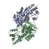
| ||||||||
|---|---|---|---|---|---|---|---|---|---|
| 1 |
| ||||||||
| Unit cell |
| ||||||||
| Atom site foot note | 1: CIS PROLINE - PRO A 138 / 2: CIS PROLINE - PRO A 195 / 3: CIS PROLINE - PRO B 138 / 4: CIS PROLINE - PRO B 195 5: THE NON-COVALENTLY BOUND COFACTOR PMP HAS BEEN GIVEN THE RESIDUE NUMBER 411 FOR CHAINS *A* AND *B*. | ||||||||
| Noncrystallographic symmetry (NCS) | NCS oper: (Code: given Matrix: (-0.92423, 0.34441, -0.16487), Vector: Details | THE TRANSFORMATION PRESENTED ON *MTRIX* RECORDS BELOW WILL YIELD APPROXIMATE COORDINATES FOR CHAIN *B* WHEN APPLIED TO CHAIN *A*, WITH AN RMSD=0.73. THE MOLECULE IS AN ALPHA2 DIMER. THE SUBUNITS ARE RELATED BY A LOCAL TWO-FOLD ROTATION AXIS. THEY ARE DISTINGUISHED BY CHAIN IDENTIFIERS A AND B, RESPECTIVELY. THE RESIDUES IN EACH SUBUNIT ARE NUMBERED FROM 3 TO 410, ACCORDING TO A SEQUENCE ALIGNMENT WITH THE LONGER SEQUENCE OF PIG CYTOSOLIC ASPARTATE AMINOTRANSFERASE (SEE U. GRAF-HAUSNER, K.J. WILSON AND P. CHRISTEN (1983). J.BIOL.CHEM. 258, 8813-8826). | |
-Components
| #1: Protein | Mass: 44992.484 Da / Num. of mol.: 2 Source method: isolated from a genetically manipulated source Source: (gene. exp.) #2: Chemical |   Mass: 248.173 Da / Num. of mol.: 2 / Source method: obtained synthetically / Formula: C8H13N2O5P Mass: 248.173 Da / Num. of mol.: 2 / Source method: obtained synthetically / Formula: C8H13N2O5P#3: Water | ChemComp-HOH / |  Mass: 18.015 Da / Num. of mol.: 620 / Source method: isolated from a natural source / Formula: H2O Mass: 18.015 Da / Num. of mol.: 620 / Source method: isolated from a natural source / Formula: H2ONonpolymer details | ONE MOLECULE OF THE COFACTOR PYRIDOXAMI | |
|---|
-Experimental details
-Experiment
| Experiment | Method: X-RAY DIFFRACTION |
|---|
- Sample preparation
| Crystal | Density Matthews: 2.33 Å3/Da / Density % sol: 47.27 % | ||||||||||||||||||||||||||||||
|---|---|---|---|---|---|---|---|---|---|---|---|---|---|---|---|---|---|---|---|---|---|---|---|---|---|---|---|---|---|---|---|
| Crystal grow | *PLUS pH: 7.5 / Method: vapor diffusion, sitting drop | ||||||||||||||||||||||||||||||
| Components of the solutions | *PLUS
|
-Data collection
| Reflection | *PLUS Highest resolution: 2.2 Å / Num. obs: 38963 |
|---|
- Processing
| Software | Name: PROLSQ / Classification: refinement | |||||||||||||||||||||||||||||||||||||||||||||||||||||||||||||||
|---|---|---|---|---|---|---|---|---|---|---|---|---|---|---|---|---|---|---|---|---|---|---|---|---|---|---|---|---|---|---|---|---|---|---|---|---|---|---|---|---|---|---|---|---|---|---|---|---|---|---|---|---|---|---|---|---|---|---|---|---|---|---|---|---|
| Refinement | Resolution: 2.2→10 Å / σ(F): 0 /
| |||||||||||||||||||||||||||||||||||||||||||||||||||||||||||||||
| Refinement step | Cycle: LAST / Resolution: 2.2→10 Å
| |||||||||||||||||||||||||||||||||||||||||||||||||||||||||||||||
| Refine LS restraints |
| |||||||||||||||||||||||||||||||||||||||||||||||||||||||||||||||
| Software | *PLUS Name: PROLSQ / Classification: refinement | |||||||||||||||||||||||||||||||||||||||||||||||||||||||||||||||
| Refinement | *PLUS Rfactor obs: 0.131 | |||||||||||||||||||||||||||||||||||||||||||||||||||||||||||||||
| Solvent computation | *PLUS | |||||||||||||||||||||||||||||||||||||||||||||||||||||||||||||||
| Displacement parameters | *PLUS Biso mean: 5.5 Å2 | |||||||||||||||||||||||||||||||||||||||||||||||||||||||||||||||
| Refine LS restraints | *PLUS
|
