 Movie
Movie Controller

[English] 日本語
 Yorodumi
- PDB-8zx8: Structure-Based Mechanism and Specificity of Human Galactosyltran... -
+ Open data

- Basic information
| Entry | Database: PDB / ID: 8zx8 | ||||||
|---|---|---|---|---|---|---|---|
| Title | Structure-Based Mechanism and Specificity of Human Galactosyltransferase B3GalT5 | ||||||
 Components Components | Beta-1,3-galactosyltransferase 5 | ||||||
 Keywords Keywords | TRANSFERASE / globo-series glycosphingolipid biosynthesis / Oxocarbenium / SNi-like / SN2-like / double displacement / inverting galactosyltransferase / Michaelis complex | ||||||
| Function / homology |  Function and homology information Function and homology informationN-acetyl-beta-D-glucosaminide beta-(1,3)-galactosyltransferase activity / Lewis blood group biosynthesis / oligosaccharide biosynthetic process / glycoprotein biosynthetic process / protein O-linked glycosylation / Transferases; Glycosyltransferases; Hexosyltransferases / response to bacterium / lipid metabolic process / Golgi membrane / endoplasmic reticulum / Golgi apparatus Similarity search - Function | ||||||
| Biological species |  Homo sapiens (human) Homo sapiens (human) | ||||||
| Method |  X-RAY DIFFRACTION / SYNCHROTRON / MOLECULAR REPLACEMENT / Resolution: 2.4 Å X-RAY DIFFRACTION / SYNCHROTRON / MOLECULAR REPLACEMENT / Resolution: 2.4 Å | ||||||
 Authors Authors | Lo, J.M. / Ma, C. | ||||||
| Funding support |  Taiwan, 1items Taiwan, 1items
| ||||||
 Citation Citation | Journal: J.Am.Chem.Soc. / Year: 2025 Title: Structure-Based Mechanism and Specificity of Human Galactosyltransferase beta 3GalT5. Authors: Lo, J.M. / Kung, C.C. / Cheng, T.R. / Wong, C.H. / Ma, C. | ||||||
| History |
|
- Structure visualization
| Structure viewer | Molecule: MolmilJmol/JSmol |
|---|
- Downloads & links
-Download
| PDBx/mmCIF format | 8zx8.cif.gz | 309.3 KB | Display | PDBx/mmCIF format |
|---|---|---|---|---|
| PDB format | pdb8zx8.ent.gz | 208.7 KB | Display | PDB format |
| PDBx/mmJSON format | 8zx8.json.gz | Tree view | PDBx/mmJSON format | |
| Others |  Other downloads Other downloads |
-Validation report
| Arichive directory | https://data.pdbj.org/pub/pdb/validation_reports/zx/8zx8ftp://data.pdbj.org/pub/pdb/validation_reports/zx/8zx8 | HTTPS FTP |
|---|
-Related structure data
| Related structure data |  8zwpC 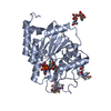 8zwrC  8zwwC  8zwyC  8zx2C  8zx3C  8zx9C C: citing same article ( |
|---|---|
| Similar structure data |
-Links
 PDBj
PDBj


- Assembly
| Deposited unit | 
| ||||||||||||
|---|---|---|---|---|---|---|---|---|---|---|---|---|---|
| 1 | 
| ||||||||||||
| 2 | 
| ||||||||||||
| Unit cell |
|
-Components
-Protein , 1 types, 2 molecules AB
| #1: Protein | Mass: 32550.551 Da / Num. of mol.: 2 Source method: isolated from a genetically manipulated source Source: (gene. exp.) Homo sapiens (human) / Gene: B3GALT5 / Cell line (production host): Sf9 / Production host:   Spodoptera frugiperda (fall armyworm) / References: UniProt: Q9Y2C3 Spodoptera frugiperda (fall armyworm) / References: UniProt: Q9Y2C3 |
|---|
-Sugars , 6 types, 7 molecules 
| #2: Polysaccharide | | #3: Polysaccharide | alpha-L-fucopyranose-(1-6)-2-acetamido-2-deoxy-beta-D-glucopyranose | #4: Polysaccharide | alpha-D-mannopyranose-(1-3)-[alpha-D-mannopyranose-(1-6)]beta-D-mannopyranose-(1-4)-2-acetamido-2- ...alpha-D-mannopyranose-(1-3)-[alpha-D-mannopyranose-(1-6)]beta-D-mannopyranose-(1-4)-2-acetamido-2-deoxy-beta-D-glucopyranose-(1-4)-[alpha-L-fucopyranose-(1-6)]2-acetamido-2-deoxy-beta-D-glucopyranose | #5: Polysaccharide | 2-acetamido-2-deoxy-beta-D-galactopyranose-(1-3)-alpha-D-galactopyranose-(1-4)-beta-D- ...2-acetamido-2-deoxy-beta-D-galactopyranose-(1-3)-alpha-D-galactopyranose-(1-4)-beta-D-galactopyranose-(1-4)-beta-D-glucopyranose | #6: Polysaccharide | 2-acetamido-2-deoxy-beta-D-galactopyranose-(1-3)-alpha-D-galactopyranose-(1-4)-beta-D-galactopyranose | Type: oligosaccharide / Mass: 545.490 Da / Num. of mol.: 1 / Source method: obtained synthetically #11: Sugar | ChemComp-GAL / |  Type: D-saccharide, beta linking / Mass: 180.156 Da / Num. of mol.: 1 / Source method: obtained synthetically / Formula: C6H12O6 / Feature type: SUBJECT OF INVESTIGATION Type: D-saccharide, beta linking / Mass: 180.156 Da / Num. of mol.: 1 / Source method: obtained synthetically / Formula: C6H12O6 / Feature type: SUBJECT OF INVESTIGATION |
|---|
-Non-polymers , 5 types, 95 molecules 


| #7: Chemical |  Mass: 54.938 Da / Num. of mol.: 2 / Source method: obtained synthetically / Formula: Mn Mass: 54.938 Da / Num. of mol.: 2 / Source method: obtained synthetically / Formula: Mn#8: Chemical |  Type: RNA linking / Mass: 404.161 Da / Num. of mol.: 2 / Source method: obtained synthetically / Formula: C9H14N2O12P2 / Comment: UDP*YM Type: RNA linking / Mass: 404.161 Da / Num. of mol.: 2 / Source method: obtained synthetically / Formula: C9H14N2O12P2 / Comment: UDP*YM#9: Chemical |  Mass: 282.334 Da / Num. of mol.: 2 / Source method: obtained synthetically / Formula: C11H26N2O6 / Comment: pH buffer*YM Mass: 282.334 Da / Num. of mol.: 2 / Source method: obtained synthetically / Formula: C11H26N2O6 / Comment: pH buffer*YM#10: Chemical |  Mass: 106.120 Da / Num. of mol.: 2 / Source method: obtained synthetically / Formula: C4H10O3 Mass: 106.120 Da / Num. of mol.: 2 / Source method: obtained synthetically / Formula: C4H10O3#12: Water | ChemComp-HOH / | Mass: 18.015 Da / Num. of mol.: 87 / Source method: isolated from a natural source / Formula: H2O |
|---|
-Details
| Has ligand of interest | Y |
|---|---|
| Has protein modification | Y |
-Experimental details
-Experiment
| Experiment | Method: X-RAY DIFFRACTION / Number of used crystals: 1 |
|---|
- Sample preparation
| Crystal | Density Matthews: 2.77 Å3/Da / Density % sol: 55.6 % |
|---|---|
| Crystal grow | Temperature: 277.15 K / Method: vapor diffusion, hanging drop Details: 0.1M BIS-TRIS Propane pH8.5, 0.2M Potassium Sodium Tartrate, 20% w/v PEG 3350 |
-Data collection
| Diffraction | Mean temperature: 100 K / Serial crystal experiment: N |
|---|---|
| Diffraction source | Source: SYNCHROTRON / Site: NSRRC / Beamline: BL15A1 / Wavelength: 1 Å |
| Detector | Type: RAYONIX MX300HE / Detector: CCD / Date: Jun 17, 2020 |
| Radiation | Protocol: SINGLE WAVELENGTH / Monochromatic (M) / Laue (L): M / Scattering type: x-ray |
| Radiation wavelength | Wavelength: 1 Å / Relative weight: 1 |
| Reflection | Resolution: 2.4→50 Å / Num. obs: 24050 / % possible obs: 98 % / Redundancy: 3.8 % / Biso Wilson estimate: 37.63 Å2 / Rmerge(I) obs: 0.13 / Net I/σ(I): 9.85 |
| Reflection shell | Resolution: 2.4→2.49 Å / Redundancy: 2.6 % / Rmerge(I) obs: 0.742 / Num. unique obs: 1042 / % possible all: 85 |
- Processing
| Software |
| |||||||||||||||||||||||||||||||||||||||||||||||||||||||||||||||
|---|---|---|---|---|---|---|---|---|---|---|---|---|---|---|---|---|---|---|---|---|---|---|---|---|---|---|---|---|---|---|---|---|---|---|---|---|---|---|---|---|---|---|---|---|---|---|---|---|---|---|---|---|---|---|---|---|---|---|---|---|---|---|---|---|
| Refinement | Method to determine structure: MOLECULAR REPLACEMENT / Resolution: 2.4→30.72 Å / SU ML: 0.3135 / Cross valid method: FREE R-VALUE / σ(F): 1.34 / Phase error: 24.8338 Stereochemistry target values: GeoStd + Monomer Library + CDL v1.2
| |||||||||||||||||||||||||||||||||||||||||||||||||||||||||||||||
| Solvent computation | Shrinkage radii: 0.9 Å / VDW probe radii: 1.11 Å / Solvent model: FLAT BULK SOLVENT MODEL | |||||||||||||||||||||||||||||||||||||||||||||||||||||||||||||||
| Displacement parameters | Biso mean: 50.11 Å2 | |||||||||||||||||||||||||||||||||||||||||||||||||||||||||||||||
| Refinement step | Cycle: LAST / Resolution: 2.4→30.72 Å
| |||||||||||||||||||||||||||||||||||||||||||||||||||||||||||||||
| Refine LS restraints |
| |||||||||||||||||||||||||||||||||||||||||||||||||||||||||||||||
| LS refinement shell |
| |||||||||||||||||||||||||||||||||||||||||||||||||||||||||||||||
| Refinement TLS params. | Method: refined / Origin x: 8.87347452071 Å / Origin y: 8.2221905263 Å / Origin z: 15.0324233144 Å
| |||||||||||||||||||||||||||||||||||||||||||||||||||||||||||||||
| Refinement TLS group | Selection details: all |
