 Movie
Movie Controller

[English] 日本語
 Yorodumi
- PDB-8zwt: Crystal structure of atypical two cysteine thiol peroxidase from ... -
+ Open data

- Basic information
| Entry | Database: PDB / ID: 8zwt | ||||||
|---|---|---|---|---|---|---|---|
| Title | Crystal structure of atypical two cysteine thiol peroxidase from Staphylococcus aureus | ||||||
 Components Components | Thiol peroxidase | ||||||
 Keywords Keywords | OXIDOREDUCTASE / Two-Cysteine Thiol peroxidase / Staphylococcus aureus / Substrate free form | ||||||
| Function / homology |  Function and homology information Function and homology information | ||||||
| Biological species |   Staphylococcus aureus (bacteria) Staphylococcus aureus (bacteria) | ||||||
| Method |  X-RAY DIFFRACTION / SYNCHROTRON / MOLECULAR REPLACEMENT / Resolution: 1.85 Å X-RAY DIFFRACTION / SYNCHROTRON / MOLECULAR REPLACEMENT / Resolution: 1.85 Å | ||||||
 Authors Authors | Shukla, M. / Maji, S. / Yadav, V.K. / Bhattacharyya, S. | ||||||
| Funding support |  India, 1items India, 1items
| ||||||
 Citation Citation | Journal: To Be Published Title: Crystal structure of atypical two cysteine thiol peroxidase from Staphylococcus aureus Authors: Shukla, M. / Maji, S. / Yadav, V.K. / Bhattacharyya, S. | ||||||
| History |
|
- Structure visualization
| Structure viewer | Molecule: MolmilJmol/JSmol |
|---|
- Downloads & links
-Download
| PDBx/mmCIF format | 8zwt.cif.gz | 245.1 KB | Display | PDBx/mmCIF format |
|---|---|---|---|---|
| PDB format | pdb8zwt.ent.gz | 165.8 KB | Display | PDB format |
| PDBx/mmJSON format | 8zwt.json.gz | Tree view | PDBx/mmJSON format | |
| Others |  Other downloads Other downloads |
-Validation report
| Arichive directory | https://data.pdbj.org/pub/pdb/validation_reports/zw/8zwtftp://data.pdbj.org/pub/pdb/validation_reports/zw/8zwt | HTTPS FTP |
|---|
-Related structure data
| Related structure data |  1psqS S: Starting model for refinement |
|---|---|
| Similar structure data |

-Links
 PDBj
PDBj

- Assembly
| Deposited unit | 
| ||||||||||||
|---|---|---|---|---|---|---|---|---|---|---|---|---|---|
| 1 |
| ||||||||||||
| Unit cell |
|
-Components
| #1: Protein | Mass: 18023.246 Da / Num. of mol.: 2 Source method: isolated from a genetically manipulated source Source: (gene. exp.) Staphylococcus aureus (bacteria) / Gene: tpx, SACOL1762 / Production host: References: UniProt: Q5HF61, thioredoxin-dependent peroxiredoxin #2: Chemical | ChemComp-DTU / ( | 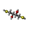  Mass: 154.251 Da / Num. of mol.: 1 / Source method: obtained synthetically / Formula: C4H10O2S2 Mass: 154.251 Da / Num. of mol.: 1 / Source method: obtained synthetically / Formula: C4H10O2S2#3: Water | ChemComp-HOH / |  Mass: 18.015 Da / Num. of mol.: 266 / Source method: isolated from a natural source / Formula: H2O Mass: 18.015 Da / Num. of mol.: 266 / Source method: isolated from a natural source / Formula: H2OHas ligand of interest | N | Has protein modification | N | |
|---|
-Experimental details
-Experiment
| Experiment | Method: X-RAY DIFFRACTION / Number of used crystals: 1 |
|---|
- Sample preparation
| Crystal | Density Matthews: 2.21 Å3/Da / Density % sol: 44.22 % |
|---|---|
| Crystal grow | Temperature: 298 K / Method: vapor diffusion, hanging drop / Details: Sodium citrate tribasic dihydrate, HEPES |
-Data collection
| Diffraction | Mean temperature: 100 K / Serial crystal experiment: N |
|---|---|
| Diffraction source | Source: SYNCHROTRON / Site: RRCAT INDUS-2 / Beamline: PX-BL21 / Wavelength: 0.97893 Å |
| Detector | Type: MAR scanner 345 mm plate / Detector: IMAGE PLATE / Date: Mar 11, 2024 |
| Radiation | Protocol: SINGLE WAVELENGTH / Monochromatic (M) / Laue (L): M / Scattering type: x-ray |
| Radiation wavelength | Wavelength: 0.97893 Å / Relative weight: 1 |
| Reflection | Resolution: 1.85→44.16 Å / Num. obs: 27499 / % possible obs: 98.4 % / Redundancy: 7 % / Biso Wilson estimate: 25.95 Å2 / CC1/2: 0.999 / Net I/σ(I): 13.9 |
| Reflection shell | Resolution: 1.85→1.95 Å / Num. unique obs: 3560 / CC1/2: 0.819 / % possible all: 89.4 |
- Processing
| Software |
| ||||||||||||||||||||||||||||||||||||||||||||||||||||||||||||||||||||||
|---|---|---|---|---|---|---|---|---|---|---|---|---|---|---|---|---|---|---|---|---|---|---|---|---|---|---|---|---|---|---|---|---|---|---|---|---|---|---|---|---|---|---|---|---|---|---|---|---|---|---|---|---|---|---|---|---|---|---|---|---|---|---|---|---|---|---|---|---|---|---|---|
| Refinement | Method to determine structure: MOLECULAR REPLACEMENT Starting model: 1PSQ Resolution: 1.85→37.25 Å / SU ML: 0.1894 / Cross valid method: FREE R-VALUE / σ(F): 1.33 / Phase error: 23.7913 Stereochemistry target values: GeoStd + Monomer Library + CDL v1.2
| ||||||||||||||||||||||||||||||||||||||||||||||||||||||||||||||||||||||
| Solvent computation | Shrinkage radii: 0.9 Å / VDW probe radii: 1.1 Å / Solvent model: FLAT BULK SOLVENT MODEL | ||||||||||||||||||||||||||||||||||||||||||||||||||||||||||||||||||||||
| Displacement parameters | Biso mean: 31.73 Å2 | ||||||||||||||||||||||||||||||||||||||||||||||||||||||||||||||||||||||
| Refinement step | Cycle: LAST / Resolution: 1.85→37.25 Å
| ||||||||||||||||||||||||||||||||||||||||||||||||||||||||||||||||||||||
| Refine LS restraints |
| ||||||||||||||||||||||||||||||||||||||||||||||||||||||||||||||||||||||
| LS refinement shell |
| ||||||||||||||||||||||||||||||||||||||||||||||||||||||||||||||||||||||
| Refinement TLS params. | Method: refined / Origin x: 0.791132926036 Å / Origin y: -5.07415783847 Å / Origin z: 12.9555276368 Å
| ||||||||||||||||||||||||||||||||||||||||||||||||||||||||||||||||||||||
| Refinement TLS group | Selection details: all |
