 Movie
Movie Controller


+ Open data

- Basic information
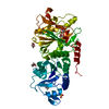
| Entry | Database: PDB / ID: 8zva | ||||||
|---|---|---|---|---|---|---|---|
| Title | structure of ShosA from E.coli APEC O1 | ||||||
 Components Components | DNA processing protein DprA | ||||||
 Keywords Keywords | IMMUNE SYSTEM / DprA / antitoxin / ShosA | ||||||
| Function / homology | DNA recombination-mediator protein A / DNA recombination-mediator protein A / DNA-mediated transformation / DNA processing protein DprA Function and homology information Function and homology information | ||||||
| Biological species |  | ||||||
| Method |  X-RAY DIFFRACTION / SYNCHROTRON / MOLECULAR REPLACEMENT / Resolution: 1.9 Å X-RAY DIFFRACTION / SYNCHROTRON / MOLECULAR REPLACEMENT / Resolution: 1.9 Å | ||||||
 Authors Authors | Yu, Y. / Chen, Q. / Pu, H. | ||||||
| Funding support | 1items
| ||||||
 Citation Citation | Journal: Nat Commun / Year: 2025 Title: A toxin-antitoxin system provides phage defense via DNA damage and repair. Authors: Pu, H. / Chen, Y. / Zhao, X. / Dai, L. / Tong, A. / Tang, D. / Chen, Q. / Yu, Y. | ||||||
| History |
|
- Structure visualization
| Structure viewer | Molecule: MolmilJmol/JSmol |
|---|
- Downloads & links
-Download
| PDBx/mmCIF format | 8zva.cif.gz | 155.6 KB | Display | PDBx/mmCIF format |
|---|---|---|---|---|
| PDB format | pdb8zva.ent.gz | 99.9 KB | Display | PDB format |
| PDBx/mmJSON format | 8zva.json.gz | Tree view | PDBx/mmJSON format | |
| Others |  Other downloads Other downloads |
-Validation report
| Summary document | 8zva_validation.pdf.gz | 443.4 KB | Display | wwPDB validaton report |
|---|---|---|---|---|
| Full document | 8zva_full_validation.pdf.gz | 448 KB | Display | |
| Data in XML | 8zva_validation.xml.gz | 15.5 KB | Display | |
| Data in CIF | 8zva_validation.cif.gz | 19.6 KB | Display | |
| Arichive directory | https://data.pdbj.org/pub/pdb/validation_reports/zv/8zvaftp://data.pdbj.org/pub/pdb/validation_reports/zv/8zva | HTTPS FTP |
-Related structure data


-Links
 PDBj
PDBj- Assembly
| Deposited unit | 
| ||||||||||||
|---|---|---|---|---|---|---|---|---|---|---|---|---|---|
| 1 | 
| ||||||||||||
| 2 | 
| ||||||||||||
| Unit cell |
| ||||||||||||
| Components on special symmetry positions |
|
-Components
| #1: Protein | Mass: 35212.969 Da / Num. of mol.: 1 Source method: isolated from a genetically manipulated source Source: (gene. exp.) Gene: APECO1_1182 / Production host: | ||||||||
|---|---|---|---|---|---|---|---|---|---|
| #2: Chemical |   Mass: 92.094 Da / Num. of mol.: 2 / Source method: obtained synthetically / Formula: C3H8O3 Mass: 92.094 Da / Num. of mol.: 2 / Source method: obtained synthetically / Formula: C3H8O3#3: Chemical | ChemComp-CL / |   Mass: 35.453 Da / Num. of mol.: 1 / Source method: obtained synthetically / Formula: Cl Mass: 35.453 Da / Num. of mol.: 1 / Source method: obtained synthetically / Formula: Cl#4: Water | ChemComp-HOH / |  Mass: 18.015 Da / Num. of mol.: 55 / Source method: isolated from a natural source / Formula: H2O Mass: 18.015 Da / Num. of mol.: 55 / Source method: isolated from a natural source / Formula: H2OHas ligand of interest | N | Has protein modification | N | |
-Experimental details
-Experiment
| Experiment | Method: X-RAY DIFFRACTION / Number of used crystals: 1 |
|---|
- Sample preparation
| Crystal | Density Matthews: 2.09 Å3/Da / Density % sol: 41.28 % |
|---|---|
| Crystal grow | Temperature: 289 K / Method: vapor diffusion, hanging drop Details: 18% PEG 2000,0.1M imidazole pH 7.0, 0.2M Ammonia citrate pH7.0 |
-Data collection
| Diffraction | Mean temperature: 100 K / Serial crystal experiment: N |
|---|---|
| Diffraction source | Source: SYNCHROTRON / Site: SSRF  / Beamline: BL19U1 / Wavelength: 0.97915 Å / Beamline: BL19U1 / Wavelength: 0.97915 Å |
| Detector | Type: DECTRIS PILATUS3 6M / Detector: PIXEL / Date: Oct 15, 2022 |
| Radiation | Protocol: SINGLE WAVELENGTH / Monochromatic (M) / Laue (L): M / Scattering type: x-ray |
| Radiation wavelength | Wavelength: 0.97915 Å / Relative weight: 1 |
| Reflection | Resolution: 1.9→50 Å / Num. obs: 24225 / % possible obs: 94.7 % / Redundancy: 20 % / Biso Wilson estimate: 30.73 Å2 / CC1/2: 1 / Net I/σ(I): 21 |
| Reflection shell | Resolution: 1.9→2.02 Å / Num. unique obs: 3239 / CC1/2: 0.945 |
- Processing
| Software |
| |||||||||||||||||||||||||||||||||||||||||||||||||||||||||||||||
|---|---|---|---|---|---|---|---|---|---|---|---|---|---|---|---|---|---|---|---|---|---|---|---|---|---|---|---|---|---|---|---|---|---|---|---|---|---|---|---|---|---|---|---|---|---|---|---|---|---|---|---|---|---|---|---|---|---|---|---|---|---|---|---|---|
| Refinement | Method to determine structure: MOLECULAR REPLACEMENT / Resolution: 1.9→49.52 Å / SU ML: 0.3945 / Cross valid method: FREE R-VALUE / σ(F): 1.33 / Phase error: 42.767 Stereochemistry target values: GeoStd + Monomer Library + CDL v1.2
| |||||||||||||||||||||||||||||||||||||||||||||||||||||||||||||||
| Solvent computation | Shrinkage radii: 0.9 Å / VDW probe radii: 1.11 Å / Solvent model: FLAT BULK SOLVENT MODEL | |||||||||||||||||||||||||||||||||||||||||||||||||||||||||||||||
| Displacement parameters | Biso mean: 51.27 Å2 | |||||||||||||||||||||||||||||||||||||||||||||||||||||||||||||||
| Refinement step | Cycle: LAST / Resolution: 1.9→49.52 Å
| |||||||||||||||||||||||||||||||||||||||||||||||||||||||||||||||
| Refine LS restraints |
| |||||||||||||||||||||||||||||||||||||||||||||||||||||||||||||||
| LS refinement shell |
|
