 Movie
Movie Controller

[English] 日本語
 Yorodumi
- PDB-8ztf: Structure of calcium preference ATP-gated channel P2X1 in the Pre... -
+ Open data

- Basic information
| Entry | Database: PDB / ID: 8ztf | |||||||||||||||||||||||||||
|---|---|---|---|---|---|---|---|---|---|---|---|---|---|---|---|---|---|---|---|---|---|---|---|---|---|---|---|---|
| Title | Structure of calcium preference ATP-gated channel P2X1 in the Pre-open state in the presence of 10mM Calcium ion | |||||||||||||||||||||||||||
 Components Components | P2X purinoceptor 1 | |||||||||||||||||||||||||||
 Keywords Keywords | TRANSPORT PROTEIN / ion channel | |||||||||||||||||||||||||||
| Function / homology |  Function and homology information Function and homology informationPlatelet homeostasis / Elevation of cytosolic Ca2+ levels / insemination / positive regulation of monoatomic ion transport / positive regulation of calcium ion import across plasma membrane / suramin binding / serotonin secretion by platelet / regulation of vascular associated smooth muscle contraction / extracellularly ATP-gated monoatomic cation channel activity / purinergic nucleotide receptor activity ...Platelet homeostasis / Elevation of cytosolic Ca2+ levels / insemination / positive regulation of monoatomic ion transport / positive regulation of calcium ion import across plasma membrane / suramin binding / serotonin secretion by platelet / regulation of vascular associated smooth muscle contraction / extracellularly ATP-gated monoatomic cation channel activity / purinergic nucleotide receptor activity / regulation of presynaptic cytosolic calcium ion concentration / regulation of smooth muscle contraction / ceramide biosynthetic process / ligand-gated calcium channel activity / regulation of synaptic vesicle exocytosis / response to ATP / regulation of calcium ion transport / monoatomic cation transmembrane transport / regulation of vasoconstriction / monoatomic cation channel activity / monoatomic ion transport / neuronal action potential / Neutrophil degranulation / presynaptic active zone membrane / synaptic transmission, glutamatergic / platelet activation / regulation of blood pressure / calcium ion transmembrane transport / monoatomic ion transmembrane transport / postsynaptic membrane / membrane raft / external side of plasma membrane / nucleotide binding / apoptotic process / protein-containing complex binding / glutamatergic synapse / protein-containing complex / ATP binding / identical protein binding / plasma membrane Similarity search - Function | |||||||||||||||||||||||||||
| Biological species |   Mus musculus (house mouse) Mus musculus (house mouse) | |||||||||||||||||||||||||||
| Method | ELECTRON MICROSCOPY / single particle reconstruction / cryo EM / Resolution: 2.43 Å | |||||||||||||||||||||||||||
 Authors Authors | Zhang, H. / Xu, H.E. | |||||||||||||||||||||||||||
| Funding support |  China, 1items China, 1items
| |||||||||||||||||||||||||||
 Citation Citation | Journal: Cell Discov / Year: 2026 Title: Structural basis for a central permeation pathway in the P2X1 receptor. Authors: Heng Zhang / Pengpeng Wu / Zhiyong Gu / Youwei Xu / Wen Hu / Qingning Yuan / Bingqing Xia / H Eric Xu / Zhaobing Gao / Abstract: The ion permeation pathway is a critical determinant of ion channel function and selectivity; however, the structural basis for ion permeation in the P2X1 receptor, an ATP-gated ion channel crucial ...The ion permeation pathway is a critical determinant of ion channel function and selectivity; however, the structural basis for ion permeation in the P2X1 receptor, an ATP-gated ion channel crucial for platelet activation, thrombosis, and male infertility, remains incompletely understood. Here, we present high-resolution cryo-electron microscopy (cryo-EM) structures of the P2X1 receptor, which reveal a central ion permeation pathway spanning the entire extracellular domain, complementing the existing paradigms of ion channel architecture for the P2X receptor family. Within this pathway, we identify specific sites that coordinate hydrated calcium ions, including an aspartate ring that acts as a selectivity filter at the apex of the central vestibule. We also discover that a small molecule, 3,5-bis(trifluoromethyl)aniline, binds at the top of the central vestibule and potently inhibits cation flux through this central permeation pathway. Our findings reveal a new inhibitor-binding site in the P2X1 receptor. These insights provide a structural framework for the rational design of subtype-specific P2X receptor inhibitors targeting the central vestibule. | |||||||||||||||||||||||||||
| History |
|
- Structure visualization
| Structure viewer | Molecule: MolmilJmol/JSmol |
|---|
- Downloads & links
-Download
| PDBx/mmCIF format | 8ztf.cif.gz | 185.5 KB | Display | PDBx/mmCIF format |
|---|---|---|---|---|
| PDB format | pdb8ztf.ent.gz | 141.2 KB | Display | PDB format |
| PDBx/mmJSON format | 8ztf.json.gz | Tree view | PDBx/mmJSON format | |
| Others |  Other downloads Other downloads |
-Validation report
| Arichive directory | https://data.pdbj.org/pub/pdb/validation_reports/zt/8ztfftp://data.pdbj.org/pub/pdb/validation_reports/zt/8ztf | HTTPS FTP |
|---|
-Related structure data
| Related structure data |  60460MC  8zt1C  8zt2C  8zt5C  8zt8C  8ztaC  8ztdC  9xqrC M: map data used to model this data C: citing same article ( |
|---|---|
| Similar structure data |


-Links
 PDBj
PDBj

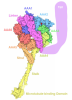
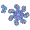
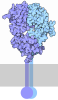
- Assembly
| Deposited unit | 
|
|---|---|
| 1 |
|
-Components
-Protein / Sugars , 2 types, 11 molecules ABC
| #1: Protein | Mass: 44908.691 Da / Num. of mol.: 3 Source method: isolated from a genetically manipulated source Source: (gene. exp.) Mus musculus (house mouse) / Gene: P2rx1 / Production host:   Spodoptera frugiperda (fall armyworm) / References: UniProt: P51576 Spodoptera frugiperda (fall armyworm) / References: UniProt: P51576#3: Sugar | ChemComp-NAG /  Type: D-saccharide, beta linking / Mass: 221.208 Da / Num. of mol.: 8 / Source method: obtained synthetically / Formula: C8H15NO6 / Feature type: SUBJECT OF INVESTIGATION Type: D-saccharide, beta linking / Mass: 221.208 Da / Num. of mol.: 8 / Source method: obtained synthetically / Formula: C8H15NO6 / Feature type: SUBJECT OF INVESTIGATION |
|---|
-Non-polymers , 4 types, 12 molecules 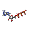



| #2: Chemical | 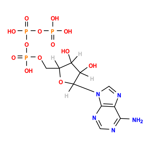 Mass: 507.181 Da / Num. of mol.: 3 / Source method: obtained synthetically / Formula: C10H16N5O13P3 / Feature type: SUBJECT OF INVESTIGATION / Comment: ATP, energy-carrying molecule*YM Mass: 507.181 Da / Num. of mol.: 3 / Source method: obtained synthetically / Formula: C10H16N5O13P3 / Feature type: SUBJECT OF INVESTIGATION / Comment: ATP, energy-carrying molecule*YM#4: Chemical |  Mass: 24.305 Da / Num. of mol.: 3 / Source method: obtained synthetically / Formula: Mg / Feature type: SUBJECT OF INVESTIGATION Mass: 24.305 Da / Num. of mol.: 3 / Source method: obtained synthetically / Formula: Mg / Feature type: SUBJECT OF INVESTIGATION#5: Chemical |  Mass: 40.078 Da / Num. of mol.: 2 / Source method: obtained synthetically / Formula: Ca / Feature type: SUBJECT OF INVESTIGATION Mass: 40.078 Da / Num. of mol.: 2 / Source method: obtained synthetically / Formula: Ca / Feature type: SUBJECT OF INVESTIGATION#6: Water | ChemComp-HOH / | Mass: 18.015 Da / Num. of mol.: 4 / Source method: isolated from a natural source / Formula: H2O |
|---|
-Details
| Has ligand of interest | Y |
|---|---|
| Has protein modification | Y |
-Experimental details
-Experiment
| Experiment | Method: ELECTRON MICROSCOPY |
|---|---|
| EM experiment | Aggregation state: PARTICLE / 3D reconstruction method: single particle reconstruction |
- Sample preparation
| Component | Name: structure of Apo p2x1 / Type: COMPLEX / Entity ID: #1 / Source: RECOMBINANT |
|---|---|
| Source (natural) | Organism: Mus musculus (house mouse) |
| Source (recombinant) | Organism: Spodoptera frugiperda (fall armyworm) |
| Buffer solution | pH: 7.3 |
| Specimen | Embedding applied: NO / Shadowing applied: NO / Staining applied: NO / Vitrification applied: YES |
| Vitrification | Cryogen name: ETHANE |
- Electron microscopy imaging
| Experimental equipment |  Model: Titan Krios / Image courtesy: FEI Company |
|---|---|
| Microscopy | Model: TFS KRIOS |
| Electron gun | Electron source:  FIELD EMISSION GUN / Accelerating voltage: 300 kV / Illumination mode: SPOT SCAN FIELD EMISSION GUN / Accelerating voltage: 300 kV / Illumination mode: SPOT SCAN |
| Electron lens | Mode: DIFFRACTION / Nominal defocus max: 3000 nm / Nominal defocus min: 1000 nm |
| Image recording | Electron dose: 50 e/Å2 / Film or detector model: FEI FALCON IV (4k x 4k) |
- Processing
| EM software | Name: PHENIX / Category: model refinement | ||||||||||||||||||||||||
|---|---|---|---|---|---|---|---|---|---|---|---|---|---|---|---|---|---|---|---|---|---|---|---|---|---|
| CTF correction | Type: PHASE FLIPPING AND AMPLITUDE CORRECTION | ||||||||||||||||||||||||
| 3D reconstruction | Resolution: 2.43 Å / Resolution method: FSC 0.143 CUT-OFF / Num. of particles: 192582 / Symmetry type: POINT | ||||||||||||||||||||||||
| Refinement | Stereochemistry target values: REAL-SPACE (WEIGHTED MAP SUM AT ATOM CENTERS) | ||||||||||||||||||||||||
| Refine LS restraints |
|
