Movie
Movie Controller


+ Open data

- Basic information
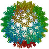
| Entry | Database: PDB / ID: 8zrh | ||||||
|---|---|---|---|---|---|---|---|
| Title | HBcAg-D4 Fab complex | ||||||
 Components Components |
| ||||||
 Keywords Keywords | VIRAL PROTEIN/IMMUNE SYSTEM / ASU / D4 Antibody / HBcAg VLP / VIRUS LIKE PARTICLE / VIRAL PROTEIN-IMMUNE SYSTEM complex | ||||||
| Function / homology |  Function and homology information Function and homology informationmicrotubule-dependent intracellular transport of viral material towards nucleus / T=4 icosahedral viral capsid / viral penetration into host nucleus / host cell / host cell cytoplasm / symbiont entry into host cell / structural molecule activity / DNA binding / RNA binding Similarity search - Function | ||||||
| Biological species |  hepatitis B virus genotype C hepatitis B virus genotype C Homo sapiens (human) Homo sapiens (human) | ||||||
| Method | ELECTRON MICROSCOPY / single particle reconstruction / cryo EM / Resolution: 3.6 Å | ||||||
 Authors Authors | Zhang, Z. / Ju, B. / Liu, C. / Yan, H. | ||||||
| Funding support |  China, 1items China, 1items
| ||||||
 Citation Citation | Journal: J Virol / Year: 2025 Title: The characterization and structural basis of a human broadly binding antibody to HBV core protein. Authors: Hu Yan / Congcong Liu / Yuxiao Li / Shilong Tang / Huimin Guo / Bing Zhou / Qing Fan / Haiyan Wang / Xiangyang Ge / Xin Wang / Xuejiao Liao / Jin Li / Zheng Zhang / Bin Ju / Abstract: Hepatitis B virus (HBV) core protein (HBc) plays a crucial role in the virus life cycle, making it an important detection marker for HBV infection and a potential target for treatment. However, ...Hepatitis B virus (HBV) core protein (HBc) plays a crucial role in the virus life cycle, making it an important detection marker for HBV infection and a potential target for treatment. However, several commercially available monoclonal antibodies (mAbs) or polyclonal antibodies (pAbs) targeting HBc have certain limitations in detecting HBV across different genotypes in various biochemical assays, such as enzyme-linked immunosorbent assay, western blot, immunofluorescence assay, flow cytometry, and immune spot assay. In this study, we identified 12 human anti-HBc mAbs and evaluated their potential application in multiple biochemical assays. These mAbs mainly recognized the epitopes near residues 20-22 amino acids (a.a.) and 77-78 a.a. of HBc, demonstrating a broadly cross-genotypic activity. Notably, three Group II mAbs, named cAbA1, cAbD4, and cAbF9, displayed excellent capacities for the detection of HBV infection in all tested biochemical assays. Furthermore, we determined a 3.22 Å of cryo-electron microscopy (cryo-EM) structure of the fragment of antigen binding (Fab) of cAbD4 complexed with HBc dimer, which was the highest resolution of the structural model for Fab-HBc to date. Collectively, our findings provided excellent and reliable antibody candidates for live HBV detection and revealed the recognition mechanism and the detailed interaction information of a potent human anti-HBc mAb binding to the spike tips of HBc dimer.IMPORTANCEThe lack of excellent detection Abs for live hepatitis B virus (HBV) infection and high-resolution structures of the Ab-HBV core protein (HBc) complex largely limited the development of HBV-related research. This study reports a panel of anti-HBc monoclonal antibodies (mAbs) with excellent capacities for detecting HBV infection in multiple biochemical assays and determines a 3.22 Å of cryo-EM structure of HBc with a potent binding mAb. These findings provide excellent and reliable detecting tools for HBV-related research and promote the understanding of the recognition mechanism of anti-HBc mAbs to HBc particles. | ||||||
| History |
|
- Structure visualization
| Structure viewer | Molecule: MolmilJmol/JSmol |
|---|
- Downloads & links
-Download
| PDBx/mmCIF format | 8zrh.cif.gz | 182.6 KB | Display | PDBx/mmCIF format |
|---|---|---|---|---|
| PDB format | pdb8zrh.ent.gz | 147.2 KB | Display | PDB format |
| PDBx/mmJSON format | 8zrh.json.gz | Tree view | PDBx/mmJSON format | |
| Others |  Other downloads Other downloads |
-Validation report
| Arichive directory | https://data.pdbj.org/pub/pdb/validation_reports/zr/8zrhftp://data.pdbj.org/pub/pdb/validation_reports/zr/8zrh | HTTPS FTP |
|---|
-Related structure data
| Related structure data | 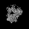 60396MC  8zreC  8zrrC C: citing same article ( M: map data used to model this data |
|---|---|
| Similar structure data |


-Links
 PDBj
PDBj


- Assembly
| Deposited unit | 
|
|---|---|
| 1 |
|
-Components
| #1: Protein | Mass: 16097.427 Da / Num. of mol.: 4 Source method: isolated from a genetically manipulated source Source: (gene. exp.) hepatitis B virus genotype C / Production host: Escherichia phage EcSzw-2 (virus) / References: UniProt: A0A679FG23#2: Antibody | Mass: 13342.837 Da / Num. of mol.: 2 Source method: isolated from a genetically manipulated source Source: (gene. exp.) Homo sapiens (human) / Production host: Homo sapiens (human)#3: Antibody | Mass: 12504.955 Da / Num. of mol.: 2 Source method: isolated from a genetically manipulated source Source: (gene. exp.) Homo sapiens (human) / Production host: Homo sapiens (human)Has protein modification | Y | |
|---|
-Experimental details
-Experiment
| Experiment | Method: ELECTRON MICROSCOPY |
|---|---|
| EM experiment | Aggregation state: PARTICLE / 3D reconstruction method: single particle reconstruction |
- Sample preparation
| Component | Name: Asymmetric unit of the complex between HBV capsid and D4 Fab Type: COMPLEX / Entity ID: #2-#3, #1 / Source: RECOMBINANT | ||||||||||||
|---|---|---|---|---|---|---|---|---|---|---|---|---|---|
| Molecular weight | Experimental value: NO | ||||||||||||
| Source (natural) |
| ||||||||||||
| Source (recombinant) |
| ||||||||||||
| Buffer solution | pH: 7.4 | ||||||||||||
| Specimen | Conc.: 3 mg/ml / Embedding applied: NO / Shadowing applied: NO / Staining applied: NO / Vitrification applied: YES | ||||||||||||
| Specimen support | Grid material: COPPER / Grid mesh size: 300 divisions/in. / Grid type: Quantifoil R1.2/1.3 | ||||||||||||
| Vitrification | Instrument: FEI VITROBOT MARK IV / Cryogen name: ETHANE / Humidity: 100 % / Chamber temperature: 277 K |
- Electron microscopy imaging
| Experimental equipment |  Model: Titan Krios / Image courtesy: FEI Company |
|---|---|
| Microscopy | Model: FEI TITAN KRIOS |
| Electron gun | Electron source:  FIELD EMISSION GUN / Accelerating voltage: 300 kV / Illumination mode: FLOOD BEAM FIELD EMISSION GUN / Accelerating voltage: 300 kV / Illumination mode: FLOOD BEAM |
| Electron lens | Mode: BRIGHT FIELD / Nominal defocus max: 2500 nm / Nominal defocus min: 1500 nm / Calibrated defocus min: 1200 nm / Calibrated defocus max: 3000 nm / Cs: 2.7 mm / C2 aperture diameter: 70 µm / Alignment procedure: COMA FREE |
| Specimen holder | Cryogen: NITROGEN / Specimen holder model: FEI TITAN KRIOS AUTOGRID HOLDER / Temperature (max): 70 K / Temperature (min): 70 K |
| Image recording | Average exposure time: 4 sec. / Electron dose: 60 e/Å2 / Film or detector model: FEI FALCON IV (4k x 4k) / Num. of grids imaged: 1 / Num. of real images: 4137 |
| Image scans | Width: 4096 / Height: 4096 |
- Processing
| EM software |
| ||||||||||||||||||||||||||||||||||||||||||||||||||
|---|---|---|---|---|---|---|---|---|---|---|---|---|---|---|---|---|---|---|---|---|---|---|---|---|---|---|---|---|---|---|---|---|---|---|---|---|---|---|---|---|---|---|---|---|---|---|---|---|---|---|---|
| CTF correction | Type: PHASE FLIPPING AND AMPLITUDE CORRECTION | ||||||||||||||||||||||||||||||||||||||||||||||||||
| Particle selection | Num. of particles selected: 817068 | ||||||||||||||||||||||||||||||||||||||||||||||||||
| 3D reconstruction | Resolution: 3.6 Å / Resolution method: FSC 0.143 CUT-OFF / Num. of particles: 193181 / Num. of class averages: 1 / Symmetry type: POINT | ||||||||||||||||||||||||||||||||||||||||||||||||||
| Atomic model building | Protocol: RIGID BODY FIT / Space: REAL / Target criteria: cross-correlation coefficient | ||||||||||||||||||||||||||||||||||||||||||||||||||
| Atomic model building | 3D fitting-ID: 1
|