 Movie
Movie Controller

[English] 日本語
 Yorodumi
- PDB-8zly: Crystal structure of Streptococcus pneumoniae pyruvate kinase in ... -
+ Open data

- Basic information
| Entry | Database: PDB / ID: 8zly | ||||||
|---|---|---|---|---|---|---|---|
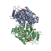
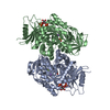
| Title | Crystal structure of Streptococcus pneumoniae pyruvate kinase in complex with oxalate and fructose 1,6-bisphosphate and UDP | ||||||
 Components Components | Pyruvate kinase | ||||||
 Keywords Keywords | TRANSFERASE / Pyruvate kinase / Streptococcus pneumoniae / Glycolysis | ||||||
| Function / homology |  Function and homology information Function and homology informationpyruvate kinase / pyruvate kinase activity / potassium ion binding / glycolytic process / kinase activity / magnesium ion binding / ATP binding / cytosol / cytoplasm Similarity search - Function | ||||||
| Biological species |  Streptococcus pneumoniae R6 (bacteria) Streptococcus pneumoniae R6 (bacteria) | ||||||
| Method |  X-RAY DIFFRACTION / SYNCHROTRON / MOLECULAR REPLACEMENT / Resolution: 1.89 Å X-RAY DIFFRACTION / SYNCHROTRON / MOLECULAR REPLACEMENT / Resolution: 1.89 Å | ||||||
 Authors Authors | Nakashima, R. / Taguchi, A. | ||||||
| Funding support |  Japan, 1items Japan, 1items
| ||||||
 Citation Citation | Journal: J.Mol.Biol. / Year: 2024 Title: Structural Basis of Nucleotide Selectivity in Pyruvate Kinase. Authors: Taguchi, A. / Nakashima, R. / Nishino, K. | ||||||
| History |
|
- Structure visualization
| Structure viewer | Molecule: MolmilJmol/JSmol |
|---|
- Downloads & links
-Download
| PDBx/mmCIF format | 8zly.cif.gz | 865 KB | Display | PDBx/mmCIF format |
|---|---|---|---|---|
| PDB format | pdb8zly.ent.gz | 700.9 KB | Display | PDB format |
| PDBx/mmJSON format | 8zly.json.gz | Tree view | PDBx/mmJSON format | |
| Others |  Other downloads Other downloads |
-Validation report
| Arichive directory | https://data.pdbj.org/pub/pdb/validation_reports/zl/8zlyftp://data.pdbj.org/pub/pdb/validation_reports/zl/8zly | HTTPS FTP |
|---|
-Related structure data

-Links
 PDBj
PDBj


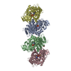
- Assembly
| Deposited unit | 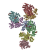
| ||||||||
|---|---|---|---|---|---|---|---|---|---|
| 1 | 
| ||||||||
| 2 | 
| ||||||||
| 3 | 
| ||||||||
| Unit cell |
|
-Components
-Protein / Sugars , 2 types, 16 molecules ABCDEFGH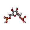
| #1: Protein | Mass: 56993.590 Da / Num. of mol.: 8 Source method: isolated from a genetically manipulated source Source: (gene. exp.) Streptococcus pneumoniae R6 (bacteria) / Gene: pykF / Production host: #2: Sugar | ChemComp-FBP /  Type: D-saccharide, beta linking / Mass: 340.116 Da / Num. of mol.: 8 / Source method: obtained synthetically / Formula: C6H14O12P2 Type: D-saccharide, beta linking / Mass: 340.116 Da / Num. of mol.: 8 / Source method: obtained synthetically / Formula: C6H14O12P2 |
|---|
-Non-polymers , 5 types, 3887 molecules 


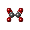

| #3: Chemical | ChemComp-MG /  Mass: 24.305 Da / Num. of mol.: 15 / Source method: obtained synthetically / Formula: Mg Mass: 24.305 Da / Num. of mol.: 15 / Source method: obtained synthetically / Formula: Mg#4: Chemical | ChemComp-UDP /  Type: RNA linking / Mass: 404.161 Da / Num. of mol.: 7 / Source method: obtained synthetically / Formula: C9H14N2O12P2 / Comment: UDP*YM Type: RNA linking / Mass: 404.161 Da / Num. of mol.: 7 / Source method: obtained synthetically / Formula: C9H14N2O12P2 / Comment: UDP*YM#5: Chemical | ChemComp-K /  Mass: 39.098 Da / Num. of mol.: 8 / Source method: obtained synthetically / Formula: K Mass: 39.098 Da / Num. of mol.: 8 / Source method: obtained synthetically / Formula: K#6: Chemical | ChemComp-OXL /  Mass: 88.019 Da / Num. of mol.: 8 / Source method: obtained synthetically / Formula: C2O4 Mass: 88.019 Da / Num. of mol.: 8 / Source method: obtained synthetically / Formula: C2O4#7: Water | ChemComp-HOH / | Mass: 18.015 Da / Num. of mol.: 3849 / Source method: isolated from a natural source / Formula: H2O |
|---|
-Details
| Has ligand of interest | N |
|---|
-Experimental details
-Experiment
| Experiment | Method: X-RAY DIFFRACTION / Number of used crystals: 1 |
|---|
- Sample preparation
| Crystal | Density Matthews: 2.91 Å3/Da / Density % sol: 57.71 % |
|---|---|
| Crystal grow | Temperature: 298 K / Method: vapor diffusion, sitting drop / pH: 7.3 Details: 60 mM HEPES pH7.3, 75 mM NaCl, 75 mM KCl, 10 mM MgCl2, 75 mM Na formate, 14 % PEG3350, 10 mM oxalate, 10 mM FBP, 5 mM UDP |
-Data collection
| Diffraction | Mean temperature: 100 K / Serial crystal experiment: N | ||||||||||||||||||||||||||||||||||||||||||||||||||||||||||||
|---|---|---|---|---|---|---|---|---|---|---|---|---|---|---|---|---|---|---|---|---|---|---|---|---|---|---|---|---|---|---|---|---|---|---|---|---|---|---|---|---|---|---|---|---|---|---|---|---|---|---|---|---|---|---|---|---|---|---|---|---|---|
| Diffraction source | Source: SYNCHROTRON / Site: SPring-8 / Beamline: BL44XU / Wavelength: 0.9 Å | ||||||||||||||||||||||||||||||||||||||||||||||||||||||||||||
| Detector | Type: DECTRIS EIGER X 16M / Detector: PIXEL / Date: Dec 4, 2023 Details: Main beamline optics is a double-crystal monochromator and a horizontal focusing mirror | ||||||||||||||||||||||||||||||||||||||||||||||||||||||||||||
| Radiation | Monochromator: Double-crystal / Protocol: SINGLE WAVELENGTH / Monochromatic (M) / Laue (L): M / Scattering type: x-ray | ||||||||||||||||||||||||||||||||||||||||||||||||||||||||||||
| Radiation wavelength | Wavelength: 0.9 Å / Relative weight: 1 | ||||||||||||||||||||||||||||||||||||||||||||||||||||||||||||
| Reflection | Resolution: 1.89→48.17 Å / Num. obs: 423736 / % possible obs: 99.8 % / Redundancy: 13.34 % / CC1/2: 0.996 / Rmerge(I) obs: 0.062 / Rrim(I) all: 0.079 / Net I/σ(I): 6.23 | ||||||||||||||||||||||||||||||||||||||||||||||||||||||||||||
| Reflection shell |
|
- Processing
| Software |
| ||||||||||||||||||||||||||||||||||||||||||||||||||||||||||||||||||||||||||||||||||||||||||||||||||||||||||||||||||||||||||||||||||||||||||||||||||||||||||||||||||||||||||||||||||||||
|---|---|---|---|---|---|---|---|---|---|---|---|---|---|---|---|---|---|---|---|---|---|---|---|---|---|---|---|---|---|---|---|---|---|---|---|---|---|---|---|---|---|---|---|---|---|---|---|---|---|---|---|---|---|---|---|---|---|---|---|---|---|---|---|---|---|---|---|---|---|---|---|---|---|---|---|---|---|---|---|---|---|---|---|---|---|---|---|---|---|---|---|---|---|---|---|---|---|---|---|---|---|---|---|---|---|---|---|---|---|---|---|---|---|---|---|---|---|---|---|---|---|---|---|---|---|---|---|---|---|---|---|---|---|---|---|---|---|---|---|---|---|---|---|---|---|---|---|---|---|---|---|---|---|---|---|---|---|---|---|---|---|---|---|---|---|---|---|---|---|---|---|---|---|---|---|---|---|---|---|---|---|---|---|
| Refinement | Method to determine structure: MOLECULAR REPLACEMENT / Resolution: 1.89→48.16 Å / Cor.coef. Fo:Fc: 0.972 / Cor.coef. Fo:Fc free: 0.958 / Cross valid method: THROUGHOUT / ESU R: 0.116 / ESU R Free: 0.116 / Stereochemistry target values: MAXIMUM LIKELIHOOD / Details: HYDROGENS HAVE BEEN ADDED IN THE RIDING POSITIONS
| ||||||||||||||||||||||||||||||||||||||||||||||||||||||||||||||||||||||||||||||||||||||||||||||||||||||||||||||||||||||||||||||||||||||||||||||||||||||||||||||||||||||||||||||||||||||
| Solvent computation | Ion probe radii: 0.8 Å / Shrinkage radii: 0.8 Å / VDW probe radii: 1.2 Å / Solvent model: MASK | ||||||||||||||||||||||||||||||||||||||||||||||||||||||||||||||||||||||||||||||||||||||||||||||||||||||||||||||||||||||||||||||||||||||||||||||||||||||||||||||||||||||||||||||||||||||
| Displacement parameters | Biso mean: 36.73 Å2
| ||||||||||||||||||||||||||||||||||||||||||||||||||||||||||||||||||||||||||||||||||||||||||||||||||||||||||||||||||||||||||||||||||||||||||||||||||||||||||||||||||||||||||||||||||||||
| Refinement step | Cycle: 1 / Resolution: 1.89→48.16 Å
| ||||||||||||||||||||||||||||||||||||||||||||||||||||||||||||||||||||||||||||||||||||||||||||||||||||||||||||||||||||||||||||||||||||||||||||||||||||||||||||||||||||||||||||||||||||||
| Refine LS restraints |
|