 Movie
Movie Controller


+ Open data

- Basic information
| Entry | Database: PDB / ID: 8zkc | ||||||
|---|---|---|---|---|---|---|---|
| Title | iron-sulfur cluster transfer protein ApbC | ||||||
 Components Components | Iron-sulfur cluster carrier protein | ||||||
 Keywords Keywords | CYTOSOLIC PROTEIN / Iron-sulfur clusters / dimerization / disulfide bonds | ||||||
| Function / homology |  Function and homology information Function and homology informationATP-dependent FeS chaperone activity / iron-sulfur cluster assembly / 4 iron, 4 sulfur cluster binding / ATP hydrolysis activity / ATP binding / metal ion binding / cytosol Similarity search - Function | ||||||
| Biological species |  | ||||||
| Method |  X-RAY DIFFRACTION / SYNCHROTRON / MOLECULAR REPLACEMENT / Resolution: 2.85 Å X-RAY DIFFRACTION / SYNCHROTRON / MOLECULAR REPLACEMENT / Resolution: 2.85 Å | ||||||
 Authors Authors | Yang, J. / Liu, L. | ||||||
| Funding support | 1items
| ||||||
 Citation Citation | Journal: Biochem.Biophys.Res.Commun. / Year: 2024 Title: Crystal structure of the iron-sulfur cluster transfer protein ApbC from Escherichia coli. Authors: Yang, J. / Duan, Y.F. / Liu, L. | ||||||
| History |
|
- Structure visualization
| Structure viewer | Molecule: MolmilJmol/JSmol |
|---|
- Downloads & links
-Download
| PDBx/mmCIF format | 8zkc.cif.gz | 145.7 KB | Display | PDBx/mmCIF format |
|---|---|---|---|---|
| PDB format | pdb8zkc.ent.gz | 112.9 KB | Display | PDB format |
| PDBx/mmJSON format | 8zkc.json.gz | Tree view | PDBx/mmJSON format | |
| Others |  Other downloads Other downloads |
-Validation report
| Arichive directory | https://data.pdbj.org/pub/pdb/validation_reports/zk/8zkcftp://data.pdbj.org/pub/pdb/validation_reports/zk/8zkc | HTTPS FTP |
|---|
-Related structure data
| Similar structure data |
|---|
-Links
 PDBj
PDBj- Assembly
| Deposited unit | 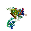
| ||||||||
|---|---|---|---|---|---|---|---|---|---|
| 1 | 
| ||||||||
| Unit cell |
|
-Components
| #1: Protein | Mass: 44206.855 Da / Num. of mol.: 1 Source method: isolated from a genetically manipulated source Source: (gene. exp.) |
|---|---|
| #2: Chemical | ChemComp-GOL /   Mass: 92.094 Da / Num. of mol.: 1 / Source method: obtained synthetically / Formula: C3H8O3 Mass: 92.094 Da / Num. of mol.: 1 / Source method: obtained synthetically / Formula: C3H8O3 |
| #3: Chemical | ChemComp-MG /   Mass: 24.305 Da / Num. of mol.: 1 / Source method: obtained synthetically / Formula: Mg Mass: 24.305 Da / Num. of mol.: 1 / Source method: obtained synthetically / Formula: Mg |
| #4: Water | ChemComp-HOH /  Mass: 18.015 Da / Num. of mol.: 1 / Source method: isolated from a natural source / Formula: H2O Mass: 18.015 Da / Num. of mol.: 1 / Source method: isolated from a natural source / Formula: H2O |
| Has ligand of interest | N |
-Experimental details
-Experiment
| Experiment | Method: X-RAY DIFFRACTION / Number of used crystals: 1 |
|---|
- Sample preparation
| Crystal | Density Matthews: 2.61 Å3/Da / Density % sol: 52.81 % |
|---|---|
| Crystal grow | Temperature: 277 K / Method: vapor diffusion, sitting drop / pH: 8 / Details: tri-potassium citrate, PEG 3350 |
-Data collection
| Diffraction | Mean temperature: 100 K / Serial crystal experiment: N |
|---|---|
| Diffraction source | Source: SYNCHROTRON / Site: SSRF  / Beamline: BL19U1 / Wavelength: 0.9795 Å / Beamline: BL19U1 / Wavelength: 0.9795 Å |
| Detector | Type: DECTRIS PILATUS3 6M / Detector: PIXEL / Date: Mar 15, 2024 |
| Radiation | Protocol: SINGLE WAVELENGTH / Monochromatic (M) / Laue (L): M / Scattering type: x-ray |
| Radiation wavelength | Wavelength: 0.9795 Å / Relative weight: 1 |
| Reflection | Resolution: 2.85→55.51 Å / Num. obs: 11707 / % possible obs: 99.9 % / Redundancy: 24.3 % / CC1/2: 1 / Rmerge(I) obs: 0.126 / Rpim(I) all: 0.026 / Rrim(I) all: 0.129 / Net I/σ(I): 21.1 |
| Reflection shell | Resolution: 2.85→2.91 Å / Mean I/σ(I) obs: 1.2 / Num. unique obs: 845 / CC1/2: 0.507 / Rpim(I) all: 0.706 |
- Processing
| Software |
| ||||||||||||||||||||||||||||||||||||||||
|---|---|---|---|---|---|---|---|---|---|---|---|---|---|---|---|---|---|---|---|---|---|---|---|---|---|---|---|---|---|---|---|---|---|---|---|---|---|---|---|---|---|
| Refinement | Method to determine structure: MOLECULAR REPLACEMENT / Resolution: 2.85→55.51 Å / SU ML: 0.39 / Cross valid method: FREE R-VALUE / σ(F): 1.36 / Phase error: 27.48 / Stereochemistry target values: ML
| ||||||||||||||||||||||||||||||||||||||||
| Solvent computation | Shrinkage radii: 0.9 Å / VDW probe radii: 1.11 Å / Solvent model: FLAT BULK SOLVENT MODEL | ||||||||||||||||||||||||||||||||||||||||
| Refinement step | Cycle: LAST / Resolution: 2.85→55.51 Å
| ||||||||||||||||||||||||||||||||||||||||
| Refine LS restraints |
| ||||||||||||||||||||||||||||||||||||||||
| LS refinement shell |
| ||||||||||||||||||||||||||||||||||||||||
| Refinement TLS params. | Method: refined / Origin x: 8.1621 Å / Origin y: 40.5851 Å / Origin z: 18.7716 Å
| ||||||||||||||||||||||||||||||||||||||||
| Refinement TLS group | Selection details: all |
