 Movie
Movie Controller


+ Open data

- Basic information
| Entry | Database: PDB / ID: 8zd3 | ||||||
|---|---|---|---|---|---|---|---|
| Title | Crystal structure of ALPK1-N+K in complex with CDP-heptose | ||||||
 Components Components | (Alpha-protein kinase ...) x 2 | ||||||
 Keywords Keywords | IMMUNE SYSTEM / complex / angonist / pattern recognition receptor | ||||||
| Function / homology |  Function and homology information Function and homology informationmonosaccharide binding / cytoplasmic pattern recognition receptor signaling pathway / cilium assembly / Alpha-protein kinase 1 signaling pathway / TAK1-dependent IKK and NF-kappa-B activation / spindle pole / positive regulation of canonical NF-kappaB signal transduction / non-specific serine/threonine protein kinase / cilium / ciliary basal body ...monosaccharide binding / cytoplasmic pattern recognition receptor signaling pathway / cilium assembly / Alpha-protein kinase 1 signaling pathway / TAK1-dependent IKK and NF-kappa-B activation / spindle pole / positive regulation of canonical NF-kappaB signal transduction / non-specific serine/threonine protein kinase / cilium / ciliary basal body / innate immune response / protein serine kinase activity / protein serine/threonine kinase activity / centrosome / ATP binding / cytosol Similarity search - Function | ||||||
| Biological species |  Homo sapiens (human) Homo sapiens (human) | ||||||
| Method |  X-RAY DIFFRACTION / SYNCHROTRON / MOLECULAR REPLACEMENT / Resolution: 2.3 Å X-RAY DIFFRACTION / SYNCHROTRON / MOLECULAR REPLACEMENT / Resolution: 2.3 Å | ||||||
 Authors Authors | She, Y. / Ding, J.J. / Shao, F. | ||||||
| Funding support | 1items
| ||||||
 Citation Citation | Journal: Science / Year: 2024 Title: The beta-d-manno-heptoses are immune agonists across kingdoms. Authors: Tang, Y. / Tian, X. / Wang, M. / Cui, Y. / She, Y. / Shi, Z. / Liu, J. / Mao, H. / Liu, L. / Li, C. / Zhang, Y. / Li, P. / Ma, Y. / Sun, J. / Du, Q. / Li, J. / Wang, J. / Li, D.F. / Wu, B. / Shao, F. / Chen, Y. | ||||||
| History |
|
- Structure visualization
| Structure viewer | Molecule: MolmilJmol/JSmol |
|---|
- Downloads & links
-Download
| PDBx/mmCIF format | 8zd3.cif.gz | 294.8 KB | Display | PDBx/mmCIF format |
|---|---|---|---|---|
| PDB format | pdb8zd3.ent.gz | Display | PDB format | |
| PDBx/mmJSON format | 8zd3.json.gz | Tree view | PDBx/mmJSON format | |
| Others |  Other downloads Other downloads |
-Validation report
| Arichive directory | https://data.pdbj.org/pub/pdb/validation_reports/zd/8zd3ftp://data.pdbj.org/pub/pdb/validation_reports/zd/8zd3 | HTTPS FTP |
|---|
-Related structure data
| Similar structure data |
|---|
-Links
 PDBj
PDBj

- Assembly
| Deposited unit | 
| ||||||||||||
|---|---|---|---|---|---|---|---|---|---|---|---|---|---|
| 1 | 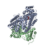
| ||||||||||||
| 2 | 
| ||||||||||||
| Unit cell |
|
-Components
-Alpha-protein kinase ... , 2 types, 4 molecules ACBD
| #1: Protein | Mass: 50672.184 Da / Num. of mol.: 2 / Fragment: N-terminal domain Source method: isolated from a genetically manipulated source Source: (gene. exp.) Homo sapiens (human) / Gene: ALPK1, KIAA1527, LAKProduction host: Insect expression vector pBlueBacmsGCB1His (others) References: UniProt: Q96QP1, non-specific serine/threonine protein kinase #2: Protein | Mass: 33570.363 Da / Num. of mol.: 2 / Fragment: C-terminal domain kinase domain Source method: isolated from a genetically manipulated source Source: (gene. exp.) Homo sapiens (human) / Gene: ALPK1, KIAA1527, LAKProduction host: Insect expression vector pBlueBacmsGCB1His (others) References: UniProt: Q96QP1, non-specific serine/threonine protein kinase |
|---|
-Sugars , 1 types, 2 molecules
| #4: Sugar | Type: D-saccharide, beta linking / Mass: 210.182 Da / Num. of mol.: 2 / Source method: obtained synthetically / Formula: C7H14O7 / Feature type: SUBJECT OF INVESTIGATION |
|---|
-Non-polymers , 3 types, 239 molecules 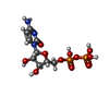


| #3: Chemical |  Mass: 403.176 Da / Num. of mol.: 2 / Source method: obtained synthetically / Formula: C9H15N3O11P2 / Feature type: SUBJECT OF INVESTIGATION Mass: 403.176 Da / Num. of mol.: 2 / Source method: obtained synthetically / Formula: C9H15N3O11P2 / Feature type: SUBJECT OF INVESTIGATION#5: Chemical |  Mass: 65.409 Da / Num. of mol.: 2 / Source method: obtained synthetically / Formula: Zn / Feature type: SUBJECT OF INVESTIGATION Mass: 65.409 Da / Num. of mol.: 2 / Source method: obtained synthetically / Formula: Zn / Feature type: SUBJECT OF INVESTIGATION#6: Water | ChemComp-HOH / | Mass: 18.015 Da / Num. of mol.: 235 / Source method: isolated from a natural source / Formula: H2O |
|---|
-Details
| Has ligand of interest | Y |
|---|---|
| Has protein modification | Y |
-Experimental details
-Experiment
| Experiment | Method: X-RAY DIFFRACTION / Number of used crystals: 1 |
|---|
- Sample preparation
| Crystal | Density Matthews: 2.35 Å3/Da / Density % sol: 47.19 % / Description: cube |
|---|---|
| Crystal grow | Temperature: 289 K / Method: vapor diffusion, hanging drop / pH: 6 / Details: 8% tacsimate ph 6.0, 14% PEG 3350 / PH range: 5.5-6.5 |
-Data collection
| Diffraction | Mean temperature: 80 K / Serial crystal experiment: N |
|---|---|
| Diffraction source | Source: SYNCHROTRON / Site: SSRF  / Beamline: BL19U1 / Wavelength: 0.97927 Å / Beamline: BL19U1 / Wavelength: 0.97927 Å |
| Detector | Type: DECTRIS PILATUS 6M / Detector: PIXEL / Date: Jul 5, 2019 |
| Radiation | Monochromator: Si(111) / Protocol: SINGLE WAVELENGTH / Monochromatic (M) / Laue (L): M / Scattering type: x-ray |
| Radiation wavelength | Wavelength: 0.97927 Å / Relative weight: 1 |
| Reflection | Resolution: 2.3→30.55 Å / Num. obs: 124422 / % possible obs: 96.39 % / Redundancy: 1.8 % / Biso Wilson estimate: 38.95 Å2 / Rmerge(I) obs: 0.041 / Net I/σ(I): 12.99 |
| Reflection shell | Resolution: 2.303→2.386 Å / Rmerge(I) obs: 0.354 / Num. unique obs: 9482 |
- Processing
| Software |
| |||||||||||||||||||||||||||||||||||||||||||||||||||||||||||||||||||||||||||||||||||||||||||||||||||||||||
|---|---|---|---|---|---|---|---|---|---|---|---|---|---|---|---|---|---|---|---|---|---|---|---|---|---|---|---|---|---|---|---|---|---|---|---|---|---|---|---|---|---|---|---|---|---|---|---|---|---|---|---|---|---|---|---|---|---|---|---|---|---|---|---|---|---|---|---|---|---|---|---|---|---|---|---|---|---|---|---|---|---|---|---|---|---|---|---|---|---|---|---|---|---|---|---|---|---|---|---|---|---|---|---|---|---|---|
| Refinement | Method to determine structure: MOLECULAR REPLACEMENT / Resolution: 2.3→30.55 Å / SU ML: 0.328 / Cross valid method: FREE R-VALUE / σ(F): 1.96 / Phase error: 31.8389 Stereochemistry target values: GeoStd + Monomer Library + CDL v1.2
| |||||||||||||||||||||||||||||||||||||||||||||||||||||||||||||||||||||||||||||||||||||||||||||||||||||||||
| Solvent computation | Shrinkage radii: 0.9 Å / VDW probe radii: 1.11 Å / Solvent model: FLAT BULK SOLVENT MODEL | |||||||||||||||||||||||||||||||||||||||||||||||||||||||||||||||||||||||||||||||||||||||||||||||||||||||||
| Displacement parameters | Biso mean: 53.1 Å2 | |||||||||||||||||||||||||||||||||||||||||||||||||||||||||||||||||||||||||||||||||||||||||||||||||||||||||
| Refinement step | Cycle: LAST / Resolution: 2.3→30.55 Å
| |||||||||||||||||||||||||||||||||||||||||||||||||||||||||||||||||||||||||||||||||||||||||||||||||||||||||
| Refine LS restraints |
| |||||||||||||||||||||||||||||||||||||||||||||||||||||||||||||||||||||||||||||||||||||||||||||||||||||||||
| LS refinement shell |
|
