[English] 日本語
 Yorodumi
- PDB-8z4l: Cryo-EM structure of CTR-bound type VII CRISPR-Cas complex at sub... -
+ Open data
- Basic information
| Entry | Database: PDB / ID: 8z4l | ||||||
|---|---|---|---|---|---|---|---|
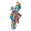
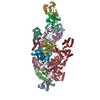
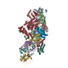
| Title | Cryo-EM structure of CTR-bound type VII CRISPR-Cas complex at substrate-engaged state I | ||||||
 Components Components |
| ||||||
 Keywords Keywords | ANTIVIRAL PROTEIN / a protein complex | ||||||
| Function / homology | RNA / RNA (> 10) Function and homology information Function and homology information | ||||||
| Biological species | metagenome (others) | ||||||
| Method | ELECTRON MICROSCOPY / single particle reconstruction / cryo EM / Resolution: 2.85 Å | ||||||
 Authors Authors | Zhang, H. / Deng, Z. / Li, X. | ||||||
| Funding support |  China, 1items China, 1items
| ||||||
 Citation Citation |  Journal: Nature / Year: 2024 Journal: Nature / Year: 2024Title: Structural basis for the activity of the type VII CRISPR-Cas system. Authors: Jie Yang / Xuzichao Li / Qiuqiu He / Xiaoshen Wang / Jingjing Tang / Tongyao Wang / Yi Zhang / Feiyang Yu / Shuqin Zhang / Zhikun Liu / Lingling Zhang / Fumeng Liao / Hang Yin / Haiyan Zhao ...Authors: Jie Yang / Xuzichao Li / Qiuqiu He / Xiaoshen Wang / Jingjing Tang / Tongyao Wang / Yi Zhang / Feiyang Yu / Shuqin Zhang / Zhikun Liu / Lingling Zhang / Fumeng Liao / Hang Yin / Haiyan Zhao / Zengqin Deng / Heng Zhang /  Abstract: The newly identified type VII CRISPR-Cas candidate system uses a CRISPR RNA-guided ribonucleoprotein complex formed by Cas5 and Cas7 proteins to target RNA. However, the RNA cleavage is executed by a ...The newly identified type VII CRISPR-Cas candidate system uses a CRISPR RNA-guided ribonucleoprotein complex formed by Cas5 and Cas7 proteins to target RNA. However, the RNA cleavage is executed by a dedicated Cas14 nuclease, which is distinct from the effector nucleases of the other CRISPR-Cas systems. Here we report seven cryo-electron microscopy structures of the Cas14-bound interference complex at different functional states. Cas14, a tetrameric protein in solution, is recruited to the Cas5-Cas7 complex in a target RNA-dependent manner. The N-terminal catalytic domain of Cas14 binds a stretch of the substrate RNA for cleavage, whereas the C-terminal domain is primarily responsible for tethering Cas14 to the Cas5-Cas7 complex. The biochemical cleavage assays corroborate the captured functional conformations, revealing that Cas14 binds to different sites on the Cas5-Cas7 complex to execute individual cleavage events. Notably, a plugged-in arginine of Cas7 sandwiched by a C-shaped clamp of C-terminal domain precisely modulates Cas14 binding. More interestingly, target RNA cleavage is altered by a complementary protospacer flanking sequence at the 5' end, but not at the 3' end. Altogether, our study elucidates critical molecular details underlying the assembly of the interference complex and substrate cleavage in the type VII CRISPR-Cas system, which may help rational engineering of the type VII CRISPR-Cas system for biotechnological applications. | ||||||
| History |
|
- Structure visualization
| Structure viewer | Molecule:  Molmil Molmil Jmol/JSmol Jmol/JSmol |
|---|
- Downloads & links
- Download
| PDBx/mmCIF format |  8z4l.cif.gz 8z4l.cif.gz | 583.9 KB | Display |  PDBx/mmCIF format PDBx/mmCIF format |
|---|---|---|---|---|
| PDB format |  pdb8z4l.ent.gz pdb8z4l.ent.gz | 462.6 KB | Display |  PDB format PDB format |
| PDBx/mmJSON format |  8z4l.json.gz 8z4l.json.gz | Tree view |  PDBx/mmJSON format PDBx/mmJSON format | |
| Others |  Other downloads Other downloads |
-Validation report
| Arichive directory |  https://data.pdbj.org/pub/pdb/validation_reports/z4/8z4l https://data.pdbj.org/pub/pdb/validation_reports/z4/8z4l ftp://data.pdbj.org/pub/pdb/validation_reports/z4/8z4l ftp://data.pdbj.org/pub/pdb/validation_reports/z4/8z4l | HTTPS FTP |
|---|
-Related structure data
| Related structure data |  39767MC  8yhdC  8yheC  8z4jC  8z99C  8z9cC  8z9eC M: map data used to model this data C: citing same article ( |
|---|---|
| Similar structure data | Similarity search - Function & homology  F&H Search F&H Search |
- Links
Links
- Assembly
| Deposited unit | 
|
|---|---|
| 1 |
|
- Components
-Protein , 3 types, 12 molecules ABCDEFJGHIKL
| #1: Protein | Mass: 22255.604 Da / Num. of mol.: 7 Source method: isolated from a genetically manipulated source Source: (gene. exp.) metagenome (others) / Production host:  #2: Protein | | Mass: 27139.533 Da / Num. of mol.: 1 Source method: isolated from a genetically manipulated source Source: (gene. exp.) metagenome (others) / Production host:  #3: Protein | Mass: 70468.898 Da / Num. of mol.: 4 Source method: isolated from a genetically manipulated source Source: (gene. exp.) metagenome (others) / Production host:  |
|---|
-RNA chain , 2 types, 2 molecules MN
| #4: RNA chain | Mass: 19142.277 Da / Num. of mol.: 1 Source method: isolated from a genetically manipulated source Source: (gene. exp.) metagenome (others) / Production host:  |
|---|---|
| #5: RNA chain | Mass: 19354.707 Da / Num. of mol.: 1 Source method: isolated from a genetically manipulated source Source: (gene. exp.) metagenome (others) / Production host:  |
-Non-polymers , 1 types, 9 molecules 
| #6: Chemical | ChemComp-ZN / |
|---|
-Details
| Has ligand of interest | N |
|---|---|
| Has protein modification | Y |
-Experimental details
-Experiment
| Experiment | Method: ELECTRON MICROSCOPY |
|---|---|
| EM experiment | Aggregation state: PARTICLE / 3D reconstruction method: single particle reconstruction |
- Sample preparation
| Component | Name: a protein / Type: COMPLEX / Entity ID: #1-#5 / Source: RECOMBINANT |
|---|---|
| Source (natural) | Organism: metagenome (others) |
| Source (recombinant) | Organism:  |
| Buffer solution | pH: 7.5 |
| Specimen | Embedding applied: NO / Shadowing applied: NO / Staining applied: NO / Vitrification applied: YES |
| Vitrification | Cryogen name: ETHANE |
- Electron microscopy imaging
| Microscopy | Model: JEOL CRYO ARM 300 |
|---|---|
| Electron gun | Electron source:  FIELD EMISSION GUN / Accelerating voltage: 300 kV / Illumination mode: FLOOD BEAM FIELD EMISSION GUN / Accelerating voltage: 300 kV / Illumination mode: FLOOD BEAM |
| Electron lens | Mode: BRIGHT FIELD / Nominal defocus max: 2500 nm / Nominal defocus min: 500 nm |
| Image recording | Electron dose: 40 e/Å2 / Film or detector model: GATAN K3 (6k x 4k) |
- Processing
| CTF correction | Type: PHASE FLIPPING ONLY |
|---|---|
| 3D reconstruction | Resolution: 2.85 Å / Resolution method: FSC 0.143 CUT-OFF / Num. of particles: 96774 / Symmetry type: POINT |
 Movie
Movie Controller


 PDBj
PDBj


