 Movie
Movie Controller

[English] 日本語
 Yorodumi
- PDB-8z2n: Crystal structure of 5-phosphomethyl-2'-deoxyuridine (5-PmdU) gly... -
+ Open data

- Basic information
| Entry | Database: PDB / ID: 8z2n | ||||||
|---|---|---|---|---|---|---|---|
| Title | Crystal structure of 5-phosphomethyl-2'-deoxyuridine (5-PmdU) glycinyltransferase gp46/PUGT from Pseudomonads phage PaMx11 in complex with dsDNA | ||||||
 Components Components |
| ||||||
 Keywords Keywords | TRANSFERASE/DNA / Pseudomonads phage / DNA / hypermodification / glycinyltransferase / 5-phosphomethyl-2'-deoxyuridine / TRANSFERASE-DNA complex | ||||||
| Function / homology | Amino acid:DNA transferase / Amino acid:DNA transferase / symbiont-mediated evasion of host restriction-modification system / transferase activity / symbiont-mediated suppression of host innate immune response / GLYCINE / DNA / DNA (> 10) / Glycinyltransferase Function and homology information Function and homology information | ||||||
| Biological species |  Pseudomonas phage PaMx11 (virus) Pseudomonas phage PaMx11 (virus)synthetic construct (others) | ||||||
| Method |  X-RAY DIFFRACTION / SYNCHROTRON / MOLECULAR REPLACEMENT / Resolution: 2.3 Å X-RAY DIFFRACTION / SYNCHROTRON / MOLECULAR REPLACEMENT / Resolution: 2.3 Å | ||||||
 Authors Authors | Wen, Y. / Guo, W.T. / Wu, B.X. | ||||||
| Funding support | 1items
| ||||||
 Citation Citation | Journal: Nucleic Acids Res. / Year: 2024 Title: Structural insights into the biosynthetic mechanism of N alpha-GlyT and 5-NmdU hypermodifications of DNA. Authors: Wen, Y. / Guo, W. / Meng, C. / Yang, J. / Xu, S. / Chen, H. / Gan, J. / Wu, B. | ||||||
| History |
|
- Structure visualization
| Structure viewer | Molecule: MolmilJmol/JSmol |
|---|
- Downloads & links
-Download
| PDBx/mmCIF format | 8z2n.cif.gz | 325.8 KB | Display | PDBx/mmCIF format |
|---|---|---|---|---|
| PDB format | pdb8z2n.ent.gz | Display | PDB format | |
| PDBx/mmJSON format | 8z2n.json.gz | Tree view | PDBx/mmJSON format | |
| Others |  Other downloads Other downloads |
-Validation report
| Arichive directory | https://data.pdbj.org/pub/pdb/validation_reports/z2/8z2nftp://data.pdbj.org/pub/pdb/validation_reports/z2/8z2n | HTTPS FTP |
|---|
-Related structure data
| Related structure data | 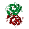 8z2mSC  8z2oC 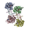 8z79C S: Starting model for refinement C: citing same article ( |
|---|---|
| Similar structure data |
-Links
 PDBj
PDBj


- Assembly
| Deposited unit | 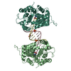
| |||||||||||||||||||||||||||||||||||||||||||||||||||||||||||||||||||||||||||||||||||||||||||||||||||||||||||
|---|---|---|---|---|---|---|---|---|---|---|---|---|---|---|---|---|---|---|---|---|---|---|---|---|---|---|---|---|---|---|---|---|---|---|---|---|---|---|---|---|---|---|---|---|---|---|---|---|---|---|---|---|---|---|---|---|---|---|---|---|---|---|---|---|---|---|---|---|---|---|---|---|---|---|---|---|---|---|---|---|---|---|---|---|---|---|---|---|---|---|---|---|---|---|---|---|---|---|---|---|---|---|---|---|---|---|---|---|
| 1 |
| |||||||||||||||||||||||||||||||||||||||||||||||||||||||||||||||||||||||||||||||||||||||||||||||||||||||||||
| Unit cell |
| |||||||||||||||||||||||||||||||||||||||||||||||||||||||||||||||||||||||||||||||||||||||||||||||||||||||||||
| Noncrystallographic symmetry (NCS) | NCS domain:
NCS domain segments:
NCS ensembles :
NCS oper:
|
-Components
-Protein , 1 types, 2 molecules BCA00
| #1: Protein | Mass: 33459.914 Da / Num. of mol.: 2 Source method: isolated from a genetically manipulated source Source: (gene. exp.) Pseudomonas phage PaMx11 (virus) / Gene: PaMx11_46 / Production host:  |
|---|
-DNA chain , 2 types, 2 molecules CD
| #2: DNA chain | Mass: 3966.597 Da / Num. of mol.: 1 / Source method: obtained synthetically / Source: (synth.) synthetic construct (others) |
|---|---|
| #3: DNA chain | Mass: 3975.611 Da / Num. of mol.: 1 / Source method: obtained synthetically / Source: (synth.) synthetic construct (others) |
-Non-polymers , 5 types, 50 molecules 


| #4: Chemical |  Mass: 96.063 Da / Num. of mol.: 2 / Source method: obtained synthetically / Formula: SO4 Mass: 96.063 Da / Num. of mol.: 2 / Source method: obtained synthetically / Formula: SO4#5: Chemical |  Mass: 40.078 Da / Num. of mol.: 2 / Source method: obtained synthetically / Formula: Ca / Feature type: SUBJECT OF INVESTIGATION Mass: 40.078 Da / Num. of mol.: 2 / Source method: obtained synthetically / Formula: Ca / Feature type: SUBJECT OF INVESTIGATION#6: Chemical | ChemComp-GOL / |  Mass: 92.094 Da / Num. of mol.: 1 / Source method: obtained synthetically / Formula: C3H8O3 Mass: 92.094 Da / Num. of mol.: 1 / Source method: obtained synthetically / Formula: C3H8O3#7: Chemical |  Type: peptide linking / Mass: 75.067 Da / Num. of mol.: 2 / Source method: obtained synthetically / Formula: C2H5NO2 Type: peptide linking / Mass: 75.067 Da / Num. of mol.: 2 / Source method: obtained synthetically / Formula: C2H5NO2#8: Water | ChemComp-HOH / | Mass: 18.015 Da / Num. of mol.: 43 / Source method: isolated from a natural source / Formula: H2O |
|---|
-Details
| Has ligand of interest | Y |
|---|---|
| Has protein modification | N |
-Experimental details
-Experiment
| Experiment | Method: X-RAY DIFFRACTION / Number of used crystals: 1 |
|---|
- Sample preparation
| Crystal | Density Matthews: 2.98 Å3/Da / Density % sol: 58.69 % |
|---|---|
| Crystal grow | Temperature: 293 K / Method: vapor diffusion, hanging drop Details: 20% (w/v) PEG 8,000, 0.1M MES/Sodium hydroxide pH 6.0, 0.2M Calcium acetate |
-Data collection
| Diffraction | Mean temperature: 100 K / Serial crystal experiment: N |
|---|---|
| Diffraction source | Source: SYNCHROTRON / Site: SSRF  / Beamline: BL02U1 / Wavelength: 0.97915 Å / Beamline: BL02U1 / Wavelength: 0.97915 Å |
| Detector | Type: DECTRIS EIGER X 16M / Detector: PIXEL / Date: Oct 15, 2022 |
| Radiation | Protocol: SINGLE WAVELENGTH / Monochromatic (M) / Laue (L): M / Scattering type: x-ray |
| Radiation wavelength | Wavelength: 0.97915 Å / Relative weight: 1 |
| Reflection | Resolution: 2.3→30 Å / Num. obs: 40612 / % possible obs: 99.9 % / Redundancy: 13.5 % / Biso Wilson estimate: 45.64 Å2 / CC1/2: 0.999 / Rmerge(I) obs: 0.154 / Rpim(I) all: 0.043 / Rrim(I) all: 0.16 / Net I/σ(I): 12.7 |
| Reflection shell | Resolution: 2.3→2.42 Å / Redundancy: 13.9 % / Rmerge(I) obs: 1.798 / Mean I/σ(I) obs: 1.5 / Num. unique obs: 5841 / CC1/2: 0.827 / Rpim(I) all: 0.496 / Rrim(I) all: 0.866 / % possible all: 99.9 |
- Processing
| Software |
| ||||||||||||||||||||||||||||||||||||||||||||||||||||||||||||||||||||||||||||||||||||||||||||||||||||||||||||||||
|---|---|---|---|---|---|---|---|---|---|---|---|---|---|---|---|---|---|---|---|---|---|---|---|---|---|---|---|---|---|---|---|---|---|---|---|---|---|---|---|---|---|---|---|---|---|---|---|---|---|---|---|---|---|---|---|---|---|---|---|---|---|---|---|---|---|---|---|---|---|---|---|---|---|---|---|---|---|---|---|---|---|---|---|---|---|---|---|---|---|---|---|---|---|---|---|---|---|---|---|---|---|---|---|---|---|---|---|---|---|---|---|---|---|
| Refinement | Method to determine structure: MOLECULAR REPLACEMENT Starting model: 8Z2M Resolution: 2.3→29 Å / SU ML: 0.3112 / Cross valid method: FREE R-VALUE / σ(F): 1.33 / Phase error: 33.488 Stereochemistry target values: GeoStd + Monomer Library + CDL v1.2
| ||||||||||||||||||||||||||||||||||||||||||||||||||||||||||||||||||||||||||||||||||||||||||||||||||||||||||||||||
| Solvent computation | Shrinkage radii: 0.9 Å / VDW probe radii: 1.1 Å / Solvent model: FLAT BULK SOLVENT MODEL | ||||||||||||||||||||||||||||||||||||||||||||||||||||||||||||||||||||||||||||||||||||||||||||||||||||||||||||||||
| Displacement parameters | Biso mean: 57.86 Å2 | ||||||||||||||||||||||||||||||||||||||||||||||||||||||||||||||||||||||||||||||||||||||||||||||||||||||||||||||||
| Refinement step | Cycle: LAST / Resolution: 2.3→29 Å
| ||||||||||||||||||||||||||||||||||||||||||||||||||||||||||||||||||||||||||||||||||||||||||||||||||||||||||||||||
| Refine LS restraints |
| ||||||||||||||||||||||||||||||||||||||||||||||||||||||||||||||||||||||||||||||||||||||||||||||||||||||||||||||||
| Refine LS restraints NCS |
| ||||||||||||||||||||||||||||||||||||||||||||||||||||||||||||||||||||||||||||||||||||||||||||||||||||||||||||||||
| LS refinement shell |
| ||||||||||||||||||||||||||||||||||||||||||||||||||||||||||||||||||||||||||||||||||||||||||||||||||||||||||||||||
| Refinement TLS params. | Method: refined / Origin x: 43.4453268248 Å / Origin y: 24.3929293907 Å / Origin z: 24.4363966068 Å
| ||||||||||||||||||||||||||||||||||||||||||||||||||||||||||||||||||||||||||||||||||||||||||||||||||||||||||||||||
| Refinement TLS group | Selection details: all |
