 Movie
Movie Controller

[English] 日本語
 Yorodumi
- PDB-8z2k: Substrate analog a013 bound form of PET-degrading cutinase mutant... -
+ Open data

- Basic information
| Entry | Database: PDB / ID: 8z2k | ||||||
|---|---|---|---|---|---|---|---|
| Title | Substrate analog a013 bound form of PET-degrading cutinase mutant Cut190**SS_S176A | ||||||
 Components Components | Alpha/beta hydrolase family protein | ||||||
 Keywords Keywords | HYDROLASE / PROTEIN ENGINEERING / POLYESTERASE / METAL BINDING / Aromatic ligand | ||||||
| Function / homology | Cutinase / PET hydrolase-like / : / carboxylic ester hydrolase activity / Alpha/Beta hydrolase fold / metal ion binding / : / Cutinase Function and homology information Function and homology information | ||||||
| Biological species |  Saccharomonospora viridis (bacteria) Saccharomonospora viridis (bacteria) | ||||||
| Method |  X-RAY DIFFRACTION / SYNCHROTRON / MOLECULAR REPLACEMENT / Resolution: 2.2 Å X-RAY DIFFRACTION / SYNCHROTRON / MOLECULAR REPLACEMENT / Resolution: 2.2 Å | ||||||
 Authors Authors | Numoto, N. / Kondo, F. / Bekker, G.J. / Liao, Z. / Yamashita, M. / Iida, A. / Ito, N. / Kamiya, N. / Oda, M. | ||||||
| Funding support |  Japan, 1items Japan, 1items
| ||||||
 Citation Citation | Journal: Int.J.Biol.Macromol. / Year: 2024 Title: Structural dynamics of the Ca 2+ -regulated cutinase towards structure-based improvement of PET degradation activity. Authors: Numoto, N. / Kondo, F. / Bekker, G.J. / Liao, Z. / Yamashita, M. / Iida, A. / Ito, N. / Kamiya, N. / Oda, M. | ||||||
| History |
|
- Structure visualization
| Structure viewer | Molecule: MolmilJmol/JSmol |
|---|
- Downloads & links
-Download
| PDBx/mmCIF format | 8z2k.cif.gz | 113.7 KB | Display | PDBx/mmCIF format |
|---|---|---|---|---|
| PDB format | pdb8z2k.ent.gz | Display | PDB format | |
| PDBx/mmJSON format | 8z2k.json.gz | Tree view | PDBx/mmJSON format | |
| Others |  Other downloads Other downloads |
-Validation report
| Arichive directory | https://data.pdbj.org/pub/pdb/validation_reports/z2/8z2kftp://data.pdbj.org/pub/pdb/validation_reports/z2/8z2k | HTTPS FTP |
|---|
-Related structure data
| Related structure data |  8z2gC  8z2hC  8z2iC 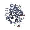 8z2jC  7ctsS S: Starting model for refinement C: citing same article ( |
|---|---|
| Similar structure data |
-Links
 PDBj
PDBj- Assembly
| Deposited unit | 
| ||||||||||||
|---|---|---|---|---|---|---|---|---|---|---|---|---|---|
| 1 | 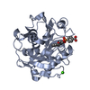
| ||||||||||||
| 2 | 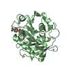
| ||||||||||||
| Unit cell |
|
-Components
| #1: Protein | Mass: 28533.098 Da / Num. of mol.: 2 / Mutation: Q123H/Q138A/S176A/N202H/S226P/R228S/D250C/E296C Source method: isolated from a genetically manipulated source Source: (gene. exp.) Saccharomonospora viridis (bacteria) / Gene: Cut190, MINT15_00360 / Plasmid: pQE80L / Production host: #2: Chemical | Mass: 336.276 Da / Num. of mol.: 2 / Source method: obtained synthetically / Formula: C16H17O6P / Feature type: SUBJECT OF INVESTIGATION #3: Chemical | ChemComp-CA / |   Mass: 40.078 Da / Num. of mol.: 1 / Source method: obtained synthetically / Formula: Ca Mass: 40.078 Da / Num. of mol.: 1 / Source method: obtained synthetically / Formula: Ca#4: Water | ChemComp-HOH / |  Mass: 18.015 Da / Num. of mol.: 80 / Source method: isolated from a natural source / Formula: H2O Mass: 18.015 Da / Num. of mol.: 80 / Source method: isolated from a natural source / Formula: H2OHas ligand of interest | Y | Has protein modification | Y | |
|---|
-Experimental details
-Experiment
| Experiment | Method: X-RAY DIFFRACTION / Number of used crystals: 1 |
|---|
- Sample preparation
| Crystal | Density Matthews: 2.33 Å3/Da / Density % sol: 47.3 % |
|---|---|
| Crystal grow | Temperature: 293 K / Method: vapor diffusion / pH: 6.8 Details: 50 mM Ammonium sulfate, 0.1 M PIPES pH 6.8, 12-18% (w/v) PEG 3,350, 0.5 mM CaCl2, and 2 mM ligands |
-Data collection
| Diffraction | Mean temperature: 100 K / Serial crystal experiment: N |
|---|---|
| Diffraction source | Source: SYNCHROTRON / Site: SPring-8 / Beamline: BL45XU / Wavelength: 0.9 Å |
| Detector | Type: DECTRIS PILATUS 6M / Detector: PIXEL / Date: May 12, 2021 |
| Radiation | Protocol: SINGLE WAVELENGTH / Monochromatic (M) / Laue (L): M / Scattering type: x-ray |
| Radiation wavelength | Wavelength: 0.9 Å / Relative weight: 1 |
| Reflection | Resolution: 2.2→49 Å / Num. obs: 26260 / % possible obs: 100 % / Redundancy: 7.9 % / CC1/2: 0.983 / Rsym value: 0.259 / Net I/σ(I): 8.3 |
| Reflection shell | Resolution: 2.2→2.28 Å / Redundancy: 7.7 % / Mean I/σ(I) obs: 1.9 / Num. unique obs: 2722 / CC1/2: 0.455 / Rsym value: 1.15 / % possible all: 100 |
- Processing
| Software |
| |||||||||||||||||||||||||||||||||||||||||||||||||||||||||||||||||||||||||||||
|---|---|---|---|---|---|---|---|---|---|---|---|---|---|---|---|---|---|---|---|---|---|---|---|---|---|---|---|---|---|---|---|---|---|---|---|---|---|---|---|---|---|---|---|---|---|---|---|---|---|---|---|---|---|---|---|---|---|---|---|---|---|---|---|---|---|---|---|---|---|---|---|---|---|---|---|---|---|---|
| Refinement | Method to determine structure: MOLECULAR REPLACEMENT Starting model: 7CTS Resolution: 2.2→48.48 Å / Cross valid method: FREE R-VALUE / σ(F): 2.78 / Phase error: 21.5169 Stereochemistry target values: GeoStd + Monomer Library + CDL v1.2
| |||||||||||||||||||||||||||||||||||||||||||||||||||||||||||||||||||||||||||||
| Solvent computation | Shrinkage radii: 0.9 Å / VDW probe radii: 1.11 Å / Solvent model: FLAT BULK SOLVENT MODEL | |||||||||||||||||||||||||||||||||||||||||||||||||||||||||||||||||||||||||||||
| Displacement parameters | Biso mean: 19.1 Å2 | |||||||||||||||||||||||||||||||||||||||||||||||||||||||||||||||||||||||||||||
| Refinement step | Cycle: LAST / Resolution: 2.2→48.48 Å
| |||||||||||||||||||||||||||||||||||||||||||||||||||||||||||||||||||||||||||||
| Refine LS restraints |
| |||||||||||||||||||||||||||||||||||||||||||||||||||||||||||||||||||||||||||||
| LS refinement shell |
|
