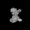
 Movie
Movie Controller


+ Open data

- Basic information
| Entry | Database: PDB / ID: 8z27 | ||||||
|---|---|---|---|---|---|---|---|
| Title | The structure of TGEV RBD and dog APN complex | ||||||
 Components Components |
| ||||||
 Keywords Keywords | ISOMERASE / TGEV / PROTEIN BINDING | ||||||
| Function / homology |  Function and homology information Function and homology informationmembrane alanyl aminopeptidase / alanyl aminopeptidase activity / peptide catabolic process / metalloaminopeptidase activity / angiotensin maturation / virus receptor activity / angiogenesis / host cell endoplasmic reticulum-Golgi intermediate compartment membrane / receptor-mediated virion attachment to host cell / cell differentiation ...membrane alanyl aminopeptidase / alanyl aminopeptidase activity / peptide catabolic process / metalloaminopeptidase activity / angiotensin maturation / virus receptor activity / angiogenesis / host cell endoplasmic reticulum-Golgi intermediate compartment membrane / receptor-mediated virion attachment to host cell / cell differentiation / endocytosis involved in viral entry into host cell / fusion of virus membrane with host plasma membrane / fusion of virus membrane with host endosome membrane / viral envelope / virion membrane / : / zinc ion binding / membrane / plasma membrane Similarity search - Function | ||||||
| Biological species |  TGEV virulent Purdue (virus) TGEV virulent Purdue (virus) | ||||||
| Method | ELECTRON MICROSCOPY / single particle reconstruction / cryo EM / Resolution: 2.86 Å | ||||||
 Authors Authors | Sun, J.Q. / Niu, S. | ||||||
| Funding support |  China, 1items China, 1items
| ||||||
 Citation Citation | Journal: PLoS Pathog / Year: 2025 Title: Cross-species recognition of two porcine coronaviruses to their cellular receptor aminopeptidase N of dogs and seven other species. Authors: Yuyang Tian / Junqing Sun / Xiaohan Hou / Zhimin Liu / Zeao Chen / Xiaoqian Pan / Ying Wang / Jianle Ren / Ding Zhang / Bo Yang / Longlong Si / Yuhai Bi / Kefang Liu / Guijun Shang / Wen-Xia ...Authors: Yuyang Tian / Junqing Sun / Xiaohan Hou / Zhimin Liu / Zeao Chen / Xiaoqian Pan / Ying Wang / Jianle Ren / Ding Zhang / Bo Yang / Longlong Si / Yuhai Bi / Kefang Liu / Guijun Shang / Wen-Xia Tian / Qihui Wang / George Fu Gao / Sheng Niu / Abstract: Porcine deltacoronavirus (PDCoV) and transmissible gastroenteritis coronavirus (TGEV), the two causative agents of porcine diarrhea, have been reported to be at risk of cross-species transmission, ...Porcine deltacoronavirus (PDCoV) and transmissible gastroenteritis coronavirus (TGEV), the two causative agents of porcine diarrhea, have been reported to be at risk of cross-species transmission, including to humans. However, the potential host range in which these two CoVs interact remains unclear. We screened 16 animal counterparts for porcine aminopeptidase N (APN), the receptor of PDCoV and TGEV, and found that APNs from eight of 17 animals could bind to the receptor-binding domains (RBDs) of PDCoV and TGEV. Furthermore, the animal APNs that could bind to the RBDs could mediate cellular infection by both viruses. Dog APN (dAPN) has been identified as the animal receptor with the highest capability to mediate the virus infection. We further resolved the complex structures of dAPN bound to the PDCoV RBD/TGEV RBD, respectively, establishing its divergent receptor-binding modes. We identified R325 of dAPN as an important residue in the PDCoV RBD-dAPN interaction, and found the central role of Q746 and T749 in dAPN in the interaction with the TGEV RBD. These findings provide the molecular basis of the potential cross-species transmission of these two porcine CoVs and shed light on future surveillance of these CoVs. | ||||||
| History |
|
- Structure visualization
| Structure viewer | Molecule: MolmilJmol/JSmol |
|---|
- Downloads & links
-Download
| PDBx/mmCIF format | 8z27.cif.gz | 397.3 KB | Display | PDBx/mmCIF format |
|---|---|---|---|---|
| PDB format | pdb8z27.ent.gz | 323 KB | Display | PDB format |
| PDBx/mmJSON format | 8z27.json.gz | Tree view | PDBx/mmJSON format | |
| Others |  Other downloads Other downloads |
-Validation report
| Arichive directory | https://data.pdbj.org/pub/pdb/validation_reports/z2/8z27ftp://data.pdbj.org/pub/pdb/validation_reports/z2/8z27 | HTTPS FTP |
|---|
-Related structure data
| Related structure data | 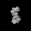 39743MC  8yziC M: map data used to model this data C: citing same article ( |
|---|---|
| Similar structure data |
-Links
 PDBj
PDBj- Assembly
| Deposited unit | 
|
|---|---|
| 1 |
|
-Components
-Protein , 2 types, 2 molecules ab
| #1: Protein | Mass: 19363.834 Da / Num. of mol.: 1 / Fragment: RBD domain Source method: isolated from a genetically manipulated source Source: (gene. exp.) TGEV virulent Purdue (virus) / Production host:  Homo sapiens (human) / References: UniProt: Q0PKZ5 Homo sapiens (human) / References: UniProt: Q0PKZ5 |
|---|---|
| #2: Protein | Mass: 107002.961 Da / Num. of mol.: 1 Source method: isolated from a genetically manipulated source Source: (gene. exp.) Homo sapiens (human) / References: UniProt: P79143, membrane alanyl aminopeptidase |
-Sugars , 4 types, 6 molecules 
| #3: Polysaccharide | beta-D-mannopyranose-(1-4)-2-acetamido-2-deoxy-beta-D-glucopyranose-(1-4)-2-acetamido-2-deoxy-beta- ...beta-D-mannopyranose-(1-4)-2-acetamido-2-deoxy-beta-D-glucopyranose-(1-4)-2-acetamido-2-deoxy-beta-D-glucopyranose Source method: isolated from a genetically manipulated source | ||||
|---|---|---|---|---|---|
| #4: Polysaccharide | Source method: isolated from a genetically manipulated source #5: Polysaccharide | beta-D-mannopyranose-(1-4)-2-acetamido-2-deoxy-beta-D-glucopyranose-(1-4)-[alpha-L-fucopyranose-(1- ...beta-D-mannopyranose-(1-4)-2-acetamido-2-deoxy-beta-D-glucopyranose-(1-4)-[alpha-L-fucopyranose-(1-6)]2-acetamido-2-deoxy-beta-D-glucopyranose | Source method: isolated from a genetically manipulated source #6: Sugar | ChemComp-NAG / |  Type: D-saccharide, beta linking / Mass: 221.208 Da / Num. of mol.: 1 / Source method: obtained synthetically / Formula: C8H15NO6 Type: D-saccharide, beta linking / Mass: 221.208 Da / Num. of mol.: 1 / Source method: obtained synthetically / Formula: C8H15NO6 |
-Details
| Has ligand of interest | N |
|---|---|
| Has protein modification | Y |
-Experimental details
-Experiment
| Experiment | Method: ELECTRON MICROSCOPY |
|---|---|
| EM experiment | Aggregation state: PARTICLE / 3D reconstruction method: single particle reconstruction |
- Sample preparation
| Component | Name: The PDCoV RBD-dAPN complex / Type: COMPLEX / Entity ID: #1-#2 / Source: RECOMBINANT |
|---|---|
| Source (natural) | Organism: |
| Source (recombinant) | Organism: Homo sapiens (human) |
| Buffer solution | pH: 8 |
| Specimen | Embedding applied: NO / Shadowing applied: NO / Staining applied: NO / Vitrification applied: YES |
| Vitrification | Cryogen name: ETHANE |
- Electron microscopy imaging
| Experimental equipment |  Model: Titan Krios / Image courtesy: FEI Company |
|---|---|
| Microscopy | Model: FEI TITAN KRIOS |
| Electron gun | Electron source:  FIELD EMISSION GUN / Accelerating voltage: 300 kV / Illumination mode: FLOOD BEAM FIELD EMISSION GUN / Accelerating voltage: 300 kV / Illumination mode: FLOOD BEAM |
| Electron lens | Mode: BRIGHT FIELD / Nominal defocus max: 2000 nm / Nominal defocus min: 1000 nm / Cs: 0.001 mm |
| Image recording | Electron dose: 60 e/Å2 / Film or detector model: GATAN K3 (6k x 4k) |
- Processing
| CTF correction | Type: PHASE FLIPPING AND AMPLITUDE CORRECTION |
|---|---|
| 3D reconstruction | Resolution: 2.86 Å / Resolution method: FSC 0.143 CUT-OFF / Num. of particles: 295620 / Symmetry type: POINT |
