[English] 日本語
 Yorodumi
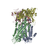
- PDB-8z1v: Cryo-EM structure of Escherichia coli DppBCDF in the resting state -
+ Open data
- Basic information
| Entry | Database: PDB / ID: 8z1v | ||||||
|---|---|---|---|---|---|---|---|
| Title | Cryo-EM structure of Escherichia coli DppBCDF in the resting state | ||||||
 Components Components |
| ||||||
 Keywords Keywords | TRANSPORT PROTEIN / ABC importer / Complex / Peptide transporter | ||||||
| Function / homology |  Function and homology information Function and homology informationABC-type dipeptide transporter / dipeptide transport / heme transmembrane transporter activity / heme transmembrane transport / dipeptide transmembrane transporter activity / ATP-binding cassette (ABC) transporter complex, substrate-binding subunit-containing / response to radiation / protein transport / 4 iron, 4 sulfur cluster binding / ATP hydrolysis activity ...ABC-type dipeptide transporter / dipeptide transport / heme transmembrane transporter activity / heme transmembrane transport / dipeptide transmembrane transporter activity / ATP-binding cassette (ABC) transporter complex, substrate-binding subunit-containing / response to radiation / protein transport / 4 iron, 4 sulfur cluster binding / ATP hydrolysis activity / ATP binding / membrane / plasma membrane Similarity search - Function | ||||||
| Biological species |  | ||||||
| Method | ELECTRON MICROSCOPY / single particle reconstruction / cryo EM / Resolution: 3.16 Å | ||||||
 Authors Authors | Li, P. / Huang, Y. | ||||||
| Funding support |  China, 1items China, 1items
| ||||||
 Citation Citation |  Journal: PLoS Biol / Year: 2025 Journal: PLoS Biol / Year: 2025Title: Structural characterization of the ABC transporter DppABCDF in Escherichia coli reveals insights into dipeptide acquisition. Authors: Panpan Li / Manfeng Zhang / Yihua Huang /  Abstract: The prokaryote-specific ATP-binding cassette (ABC) peptide transporters are involved in various physiological processes and plays an important role in transporting naturally occurring antibiotics ...The prokaryote-specific ATP-binding cassette (ABC) peptide transporters are involved in various physiological processes and plays an important role in transporting naturally occurring antibiotics across the membrane to their intracellular targets. The dipeptide transporter DppABCDF in Gram-negative bacteria is composed of five distinct subunits, yet its assembly and underlying peptide import mechanism remain elusive. Here, we report the cryo-EM structures of the DppBCDF translocator from Escherichia coli in both its apo form and in complexes bound to nonhydrolyzable or slowly hydrolyzable ATP analogs (AMPPNP and ATPγS), as well as the ATPγS-bound DppABCDF full transporter. Unlike the reported heterotrimeric Mycobacterium tuberculosis DppBCD translocator, the E. coli DppBCDF translocator is a heterotetramer, with a [4Fe-4S] cluster at the C-terminus of each ATPase subunit. Structural studies reveal that ATPγS/AMPPNP-bound DppBCDF adopts an inward-facing conformation, similar to that of apo-DppBCDF, with only one ATPγS or AMPPNP molecule bound to DppF. By contrast, ATPγS-bound DppABCDF adopts an outward-facing conformation, with two ATPγS molecules glueing DppD and DppF at the interface. Consistent with structural observations, ATPase activity assays show that the DppBCDF translocator itself is inactive and its activation requires concurrent binding of DppA and ATP. In addition, bacterial complementation experiments imply that a unique periplasmic scoop motif in DppB may play important roles in ensuring dipeptide substrates import across the membrane, presumably by preventing dipeptide back-and-forth binding to DppA and avoiding dipeptides escaping into the periplasm upon being released from DppA. | ||||||
| History |
|
- Structure visualization
| Structure viewer | Molecule:  Molmil Molmil Jmol/JSmol Jmol/JSmol |
|---|
- Downloads & links
- Download
| PDBx/mmCIF format |  8z1v.cif.gz 8z1v.cif.gz | 246.3 KB | Display |  PDBx/mmCIF format PDBx/mmCIF format |
|---|---|---|---|---|
| PDB format |  pdb8z1v.ent.gz pdb8z1v.ent.gz | 195.1 KB | Display |  PDB format PDB format |
| PDBx/mmJSON format |  8z1v.json.gz 8z1v.json.gz | Tree view |  PDBx/mmJSON format PDBx/mmJSON format | |
| Others |  Other downloads Other downloads |
-Validation report
| Summary document |  8z1v_validation.pdf.gz 8z1v_validation.pdf.gz | 1 MB | Display |  wwPDB validaton report wwPDB validaton report |
|---|---|---|---|---|
| Full document |  8z1v_full_validation.pdf.gz 8z1v_full_validation.pdf.gz | 1 MB | Display | |
| Data in XML |  8z1v_validation.xml.gz 8z1v_validation.xml.gz | 41.9 KB | Display | |
| Data in CIF |  8z1v_validation.cif.gz 8z1v_validation.cif.gz | 63.5 KB | Display | |
| Arichive directory |  https://data.pdbj.org/pub/pdb/validation_reports/z1/8z1v https://data.pdbj.org/pub/pdb/validation_reports/z1/8z1v ftp://data.pdbj.org/pub/pdb/validation_reports/z1/8z1v ftp://data.pdbj.org/pub/pdb/validation_reports/z1/8z1v | HTTPS FTP |
-Related structure data
| Related structure data |  39737MC  8z1wC  8z1xC  8z1yC M: map data used to model this data C: citing same article ( |
|---|---|
| Similar structure data | Similarity search - Function & homology  F&H Search F&H Search |
- Links
Links
- Assembly
| Deposited unit | 
|
|---|---|
| 1 |
|
- Components
| #1: Protein | Mass: 37531.812 Da / Num. of mol.: 1 Source method: isolated from a genetically manipulated source Source: (gene. exp.)   | ||||
|---|---|---|---|---|---|
| #2: Protein | Mass: 32328.295 Da / Num. of mol.: 1 Source method: isolated from a genetically manipulated source Source: (gene. exp.)   | ||||
| #3: Protein | Mass: 35887.348 Da / Num. of mol.: 1 Source method: isolated from a genetically manipulated source Source: (gene. exp.)   | ||||
| #4: Protein | Mass: 37611.438 Da / Num. of mol.: 1 Source method: isolated from a genetically manipulated source Source: (gene. exp.)   | ||||
| #5: Chemical | | Has ligand of interest | Y | Has protein modification | N | |
-Experimental details
-Experiment
| Experiment | Method: ELECTRON MICROSCOPY |
|---|---|
| EM experiment | Aggregation state: PARTICLE / 3D reconstruction method: single particle reconstruction |
- Sample preparation
| Component | Name: DppBCDF / Type: COMPLEX / Entity ID: #1-#4 / Source: RECOMBINANT |
|---|---|
| Molecular weight | Value: 0.143 MDa / Experimental value: YES |
| Source (natural) | Organism:  |
| Source (recombinant) | Organism:  |
| Buffer solution | pH: 7.5 |
| Specimen | Embedding applied: NO / Shadowing applied: NO / Staining applied: NO / Vitrification applied: YES |
| Vitrification | Cryogen name: ETHANE |
- Electron microscopy imaging
| Experimental equipment |  Model: Titan Krios / Image courtesy: FEI Company |
|---|---|
| Microscopy | Model: TFS KRIOS |
| Electron gun | Electron source:  FIELD EMISSION GUN / Accelerating voltage: 300 kV / Illumination mode: OTHER FIELD EMISSION GUN / Accelerating voltage: 300 kV / Illumination mode: OTHER |
| Electron lens | Mode: BRIGHT FIELD / Nominal defocus max: 2500 nm / Nominal defocus min: 1500 nm |
| Image recording | Electron dose: 60 e/Å2 / Film or detector model: GATAN K2 SUMMIT (4k x 4k) |
- Processing
| CTF correction | Type: PHASE FLIPPING AND AMPLITUDE CORRECTION |
|---|---|
| 3D reconstruction | Resolution: 3.16 Å / Resolution method: FSC 0.143 CUT-OFF / Num. of particles: 411381 / Symmetry type: POINT |
 Movie
Movie Controller


 PDBj
PDBj


