 Movie
Movie Controller


+ Open data

- Basic information
| Entry | Database: PDB / ID: 8z03 | ||||||||||||
|---|---|---|---|---|---|---|---|---|---|---|---|---|---|
| Title | Lactate bound to human GTP-specific succinyl-CoA synthetase | ||||||||||||
 Components Components | (Succinate--CoA ligase ...) x 2 | ||||||||||||
 Keywords Keywords | LIGASE / GTP-specific succinyl-CoA synthetase / GTPSCS / lactyl-CoA ligase | ||||||||||||
| Function / homology |  Function and homology information Function and homology informationsuccinate-CoA ligase activity / malate-CoA ligase / succinate-CoA ligase complex (GDP-forming) / succinate-CoA ligase (GDP-forming) / succinate-CoA ligase (GDP-forming) activity / succinate-CoA ligase complex (ADP-forming) / succinate-CoA ligase (ADP-forming) / succinate-CoA ligase complex / Ligases; Forming carbon-sulfur bonds; Acid-thiol ligases / succinate-CoA ligase (ADP-forming) activity ...succinate-CoA ligase activity / malate-CoA ligase / succinate-CoA ligase complex (GDP-forming) / succinate-CoA ligase (GDP-forming) / succinate-CoA ligase (GDP-forming) activity / succinate-CoA ligase complex (ADP-forming) / succinate-CoA ligase (ADP-forming) / succinate-CoA ligase complex / Ligases; Forming carbon-sulfur bonds; Acid-thiol ligases / succinate-CoA ligase (ADP-forming) activity / succinyl-CoA catabolic process / succinyl-CoA metabolic process / succinate metabolic process / Citric acid cycle (TCA cycle) / tricarboxylic acid cycle / Mitochondrial protein degradation / GDP binding / mitochondrial matrix / nucleotide binding / GTP binding / protein-containing complex binding / magnesium ion binding / mitochondrion / RNA binding / ATP binding / plasma membrane Similarity search - Function | ||||||||||||
| Biological species |  Homo sapiens (human) Homo sapiens (human) | ||||||||||||
| Method |  X-RAY DIFFRACTION / SYNCHROTRON / MOLECULAR REPLACEMENT / Resolution: 1.99 Å X-RAY DIFFRACTION / SYNCHROTRON / MOLECULAR REPLACEMENT / Resolution: 1.99 Å | ||||||||||||
 Authors Authors | Liu, R.L. / Ren, X.L. / Li, L.T. / Zhang, Y. / Huang, H. / Zhao, Y.M. | ||||||||||||
| Funding support |  United States, United States,  China, 3items China, 3items
| ||||||||||||
 Citation Citation | Journal: Cell Metab. / Year: 2025 Title: Nuclear GTPSCS functions as a lactyl-CoA synthetase to promote histone lactylation and gliomagenesis. Authors: Liu, R. / Ren, X. / Park, Y.E. / Feng, H. / Sheng, X. / Song, X. / AminiTabrizi, R. / Shah, H. / Li, L. / Zhang, Y. / Abdullah, K.G. / Dubois-Coyne, S. / Lin, H. / Cole, P.A. / DeBerardinis, ...Authors: Liu, R. / Ren, X. / Park, Y.E. / Feng, H. / Sheng, X. / Song, X. / AminiTabrizi, R. / Shah, H. / Li, L. / Zhang, Y. / Abdullah, K.G. / Dubois-Coyne, S. / Lin, H. / Cole, P.A. / DeBerardinis, R.J. / McBrayer, S.K. / Huang, H. / Zhao, Y. | ||||||||||||
| History |
|
- Structure visualization
| Structure viewer | Molecule: MolmilJmol/JSmol |
|---|
- Downloads & links
-Download
| PDBx/mmCIF format | 8z03.cif.gz | 151 KB | Display | PDBx/mmCIF format |
|---|---|---|---|---|
| PDB format | pdb8z03.ent.gz | 114.1 KB | Display | PDB format |
| PDBx/mmJSON format | 8z03.json.gz | Tree view | PDBx/mmJSON format | |
| Others |  Other downloads Other downloads |
-Validation report
| Arichive directory | https://data.pdbj.org/pub/pdb/validation_reports/z0/8z03ftp://data.pdbj.org/pub/pdb/validation_reports/z0/8z03 | HTTPS FTP |
|---|
-Related structure data
| Related structure data |  8z02C 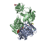 5caeS S: Starting model for refinement C: citing same article ( |
|---|---|
| Similar structure data |
-Links
 PDBj
PDBj


- Assembly
| Deposited unit | 
| ||||||||
|---|---|---|---|---|---|---|---|---|---|
| 1 |
| ||||||||
| Unit cell |
|
-Components
-Succinate--CoA ligase ... , 2 types, 2 molecules AB
| #1: Protein | Mass: 34250.133 Da / Num. of mol.: 1 Source method: isolated from a genetically manipulated source Source: (gene. exp.) Homo sapiens (human) / Gene: SUCLG1 / Production host:  References: UniProt: P53597, succinate-CoA ligase (GDP-forming), succinate-CoA ligase (ADP-forming) |
|---|---|
| #2: Protein | Mass: 42627.781 Da / Num. of mol.: 1 Source method: isolated from a genetically manipulated source Source: (gene. exp.) Homo sapiens (human) / Gene: SUCLG2 / Production host: References: UniProt: Q96I99, succinate-CoA ligase (GDP-forming) |
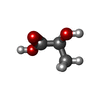
-Non-polymers , 5 types, 218 molecules 


| #3: Chemical | ChemComp-COA /  Mass: 767.534 Da / Num. of mol.: 1 / Source method: obtained synthetically / Formula: C21H36N7O16P3S / Feature type: SUBJECT OF INVESTIGATION Mass: 767.534 Da / Num. of mol.: 1 / Source method: obtained synthetically / Formula: C21H36N7O16P3S / Feature type: SUBJECT OF INVESTIGATION |
|---|---|
| #4: Chemical | ChemComp-PO4 /  Mass: 94.971 Da / Num. of mol.: 1 / Source method: obtained synthetically / Formula: PO4 / Feature type: SUBJECT OF INVESTIGATION Mass: 94.971 Da / Num. of mol.: 1 / Source method: obtained synthetically / Formula: PO4 / Feature type: SUBJECT OF INVESTIGATION |
| #5: Chemical | ChemComp-2OP / ( Mass: 90.078 Da / Num. of mol.: 1 / Source method: obtained synthetically / Formula: C3H6O3 / Feature type: SUBJECT OF INVESTIGATION Mass: 90.078 Da / Num. of mol.: 1 / Source method: obtained synthetically / Formula: C3H6O3 / Feature type: SUBJECT OF INVESTIGATION |
| #6: Chemical | ChemComp-MG /  Mass: 24.305 Da / Num. of mol.: 1 / Source method: obtained synthetically / Formula: Mg / Feature type: SUBJECT OF INVESTIGATION Mass: 24.305 Da / Num. of mol.: 1 / Source method: obtained synthetically / Formula: Mg / Feature type: SUBJECT OF INVESTIGATION |
| #7: Water | ChemComp-HOH / Mass: 18.015 Da / Num. of mol.: 214 / Source method: isolated from a natural source / Formula: H2O |
-Details
| Has ligand of interest | Y |
|---|---|
| Has protein modification | N |
-Experimental details
-Experiment
| Experiment | Method: X-RAY DIFFRACTION / Number of used crystals: 1 |
|---|
- Sample preparation
| Crystal | Density Matthews: 2.73 Å3/Da / Density % sol: 55 % |
|---|---|
| Crystal grow | Temperature: 291.15 K / Method: vapor diffusion, sitting drop Details: 0.2 M Ammonium sulfate, 0.1 M TRIS pH=7.5, 20% w/v PEG MME 5000 |
-Data collection
| Diffraction | Mean temperature: 100 K / Serial crystal experiment: N | ||||||||||||||||||||||||||||||
|---|---|---|---|---|---|---|---|---|---|---|---|---|---|---|---|---|---|---|---|---|---|---|---|---|---|---|---|---|---|---|---|
| Diffraction source | Source: SYNCHROTRON / Site: SSRF / Beamline: BL18U1 / Wavelength: 0.97915 Å | ||||||||||||||||||||||||||||||
| Detector | Type: DECTRIS PILATUS 6M / Detector: PIXEL / Date: Dec 26, 2020 | ||||||||||||||||||||||||||||||
| Radiation | Protocol: SINGLE WAVELENGTH / Monochromatic (M) / Laue (L): M / Scattering type: x-ray | ||||||||||||||||||||||||||||||
| Radiation wavelength | Wavelength: 0.97915 Å / Relative weight: 1 | ||||||||||||||||||||||||||||||
| Reflection | Resolution: 1.99→43.81 Å / Num. obs: 55815 / % possible obs: 99 % / Redundancy: 5.1 % / CC1/2: 0.986 / Rmerge(I) obs: 0.18 / Rpim(I) all: 0.087 / Rrim(I) all: 0.201 / Net I/σ(I): 6.6 / Num. measured all: 286049 / Scaling rejects: 432 | ||||||||||||||||||||||||||||||
| Reflection shell | Diffraction-ID: 1
|
- Processing
| Software |
| ||||||||||||||||||||||||||||||||||||||||||||||||||||||||||||||||||||||||||||||||||||||||||||||||||||||||||||||||||||||||||||||
|---|---|---|---|---|---|---|---|---|---|---|---|---|---|---|---|---|---|---|---|---|---|---|---|---|---|---|---|---|---|---|---|---|---|---|---|---|---|---|---|---|---|---|---|---|---|---|---|---|---|---|---|---|---|---|---|---|---|---|---|---|---|---|---|---|---|---|---|---|---|---|---|---|---|---|---|---|---|---|---|---|---|---|---|---|---|---|---|---|---|---|---|---|---|---|---|---|---|---|---|---|---|---|---|---|---|---|---|---|---|---|---|---|---|---|---|---|---|---|---|---|---|---|---|---|---|---|---|
| Refinement | Method to determine structure: MOLECULAR REPLACEMENT Starting model: 5cae Resolution: 1.99→42.129 Å / SU ML: 0.26 / Cross valid method: THROUGHOUT / σ(F): 1.39 / Phase error: 24.05 / Stereochemistry target values: ML
| ||||||||||||||||||||||||||||||||||||||||||||||||||||||||||||||||||||||||||||||||||||||||||||||||||||||||||||||||||||||||||||||
| Solvent computation | Shrinkage radii: 0.9 Å / VDW probe radii: 1.11 Å / Solvent model: FLAT BULK SOLVENT MODEL | ||||||||||||||||||||||||||||||||||||||||||||||||||||||||||||||||||||||||||||||||||||||||||||||||||||||||||||||||||||||||||||||
| Displacement parameters | Biso max: 60.55 Å2 / Biso mean: 23.8282 Å2 / Biso min: 10.46 Å2 | ||||||||||||||||||||||||||||||||||||||||||||||||||||||||||||||||||||||||||||||||||||||||||||||||||||||||||||||||||||||||||||||
| Refinement step | Cycle: final / Resolution: 1.99→42.129 Å
| ||||||||||||||||||||||||||||||||||||||||||||||||||||||||||||||||||||||||||||||||||||||||||||||||||||||||||||||||||||||||||||||
| LS refinement shell | Refine-ID: X-RAY DIFFRACTION / Rfactor Rfree error: 0
|
