 Movie
Movie Controller


+ Open data

- Basic information
| Entry | Database: PDB / ID: 8yw2 | |||||||||||||||||||||
|---|---|---|---|---|---|---|---|---|---|---|---|---|---|---|---|---|---|---|---|---|---|---|
| Title | Semliki Forest virus viron in complex with VLDLR | |||||||||||||||||||||
 Components Components |
| |||||||||||||||||||||
 Keywords Keywords | VIRAL PROTEIN / Semliki Forest virus viron / VLDLR | |||||||||||||||||||||
| Function / homology |  Function and homology information Function and homology informationreelin receptor activity / VLDL clearance / glycoprotein transport / very-low-density lipoprotein particle receptor activity / Reelin signalling pathway / ventral spinal cord development / very-low-density lipoprotein particle binding / low-density lipoprotein particle receptor activity / very-low-density lipoprotein particle clearance / reelin-mediated signaling pathway ...reelin receptor activity / VLDL clearance / glycoprotein transport / very-low-density lipoprotein particle receptor activity / Reelin signalling pathway / ventral spinal cord development / very-low-density lipoprotein particle binding / low-density lipoprotein particle receptor activity / very-low-density lipoprotein particle clearance / reelin-mediated signaling pathway / very-low-density lipoprotein particle / positive regulation of dendrite development / cargo receptor activity / lipid transport / dendrite morphogenesis / regulation of synapse assembly / apolipoprotein binding / cholesterol metabolic process / clathrin-coated pit / receptor-mediated endocytosis / VLDLR internalisation and degradation / memory / calcium-dependent protein binding / nervous system development / receptor complex / lysosomal membrane / calcium ion binding / glutamatergic synapse / signal transduction / membrane / plasma membrane Similarity search - Function | |||||||||||||||||||||
| Biological species |  Homo sapiens (human) Homo sapiens (human) Semliki Forest virus 4 Semliki Forest virus 4 | |||||||||||||||||||||
| Method | ELECTRON MICROSCOPY / single particle reconstruction / cryo EM / Resolution: 3.7 Å | |||||||||||||||||||||
 Authors Authors | Wang, J. / Zheng, T. / Yang, D. | |||||||||||||||||||||
| Funding support | 1items
| |||||||||||||||||||||
 Citation Citation | Journal: PLoS Pathog / Year: 2024 Title: Structural insights into Semiliki forest virus receptor binding modes indicate novel mechanism of virus endocytosis. Authors: Decheng Yang / Nan Wang / Bingchen Du / Zhenzhao Sun / Shida Wang / Xijun He / Jinyue Wang / Tao Zheng / Yutao Chen / Xiangxi Wang / Jingfei Wang /  Abstract: The Very Low-Density Lipoprotein Receptor (VLDLR) is an entry receptor for the prototypic alphavirus Semliki Forest Virus (SFV). However, the precise mechanisms underlying the entry of SFV into cells ...The Very Low-Density Lipoprotein Receptor (VLDLR) is an entry receptor for the prototypic alphavirus Semliki Forest Virus (SFV). However, the precise mechanisms underlying the entry of SFV into cells mediated by VLDLR remain unclear. In this study, we found that of the eight class A (LA) repeats of the VLDLR, only LA2, LA3, and LA5 specifically bind to the native SFV virion while synergistically promoting SFV cell attachment and entry. Furthermore, the multiple cryo-electron microscopy structures of VLDLR-SFV complexes and mutagenesis studies have demonstrated that under physiological conditions, VLDLR primarily binds to E1-DIII of site-1, site-2, and site-1' at the twofold symmetry axes of SFV virion through LA2, LA3, and LA5, respectively. These findings unveil a novel mechanism for viral entry mediated by receptors, suggesting that conformational transitions in VLDLR induced by multivalent binding of LAs facilitate cellular internalization of SFV, with significant implications for the design of antiviral therapeutics. | |||||||||||||||||||||
| History |
|
- Structure visualization
| Structure viewer | Molecule: MolmilJmol/JSmol |
|---|
- Downloads & links
-Download
| PDBx/mmCIF format | 8yw2.cif.gz | 2.8 MB | Display | PDBx/mmCIF format |
|---|---|---|---|---|
| PDB format | pdb8yw2.ent.gz | Display | PDB format | |
| PDBx/mmJSON format | 8yw2.json.gz | Tree view | PDBx/mmJSON format | |
| Others |  Other downloads Other downloads |
-Validation report
| Arichive directory | https://data.pdbj.org/pub/pdb/validation_reports/yw/8yw2ftp://data.pdbj.org/pub/pdb/validation_reports/yw/8yw2 | HTTPS FTP |
|---|
-Related structure data
| Related structure data |  39620MC 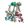 8yvyC  8yvzC  8yw0C  8yw1C M: map data used to model this data C: citing same article ( |
|---|---|
| Similar structure data |


-Links
 PDBj
PDBj


- Assembly
| Deposited unit | 
|
|---|---|
| 1 |
|
-Components
-Spike glycoprotein ... , 3 types, 48 molecules 02AAABLMNSabcgluvw456EFGHUVWhimo...
| #1: Protein | Mass: 5848.729 Da / Num. of mol.: 16 / Source method: isolated from a natural source / Source: (natural) Semliki Forest virus 4 / References: UniProt: A0A0E3T652#3: Protein | Mass: 47489.766 Da / Num. of mol.: 16 / Source method: isolated from a natural source / Source: (natural) Semliki Forest virus 4 / References: UniProt: A0A0E3T652#4: Protein | Mass: 46468.867 Da / Num. of mol.: 16 / Source method: isolated from a natural source Details: Molecular name is based on https://www.ebi.ac.uk/interpro/protein/UniProt/A0A0E3T652/ as Uniprot Reference A0A0E3T652 and author provided name are both Structural polyprotein. Source: (natural) Semliki Forest virus 4 / References: UniProt: A0A0E3T652 |
|---|
-Protein , 2 types, 17 molecules 13ACADAEDOPQTdefnxyA
| #2: Protein | Mass: 17791.299 Da / Num. of mol.: 16 / Source method: isolated from a natural source / Source: (natural) Semliki Forest virus 4 / References: UniProt: A0A0E3T652#5: Protein | | Mass: 17585.789 Da / Num. of mol.: 1 Source method: isolated from a genetically manipulated source Source: (gene. exp.) Homo sapiens (human) / Gene: VLDLR / Production host: Homo sapiens (human) / References: UniProt: P98155 |
|---|
-Sugars , 1 types, 48 molecules
| #6: Polysaccharide | beta-D-mannopyranose-(1-4)-2-acetamido-2-deoxy-beta-D-glucopyranose-(1-4)-2-acetamido-2-deoxy-beta- ...beta-D-mannopyranose-(1-4)-2-acetamido-2-deoxy-beta-D-glucopyranose-(1-4)-2-acetamido-2-deoxy-beta-D-glucopyranose Source method: isolated from a genetically manipulated source |
|---|
-Details
| Has ligand of interest | N |
|---|---|
| Has protein modification | Y |
-Experimental details
-Experiment
| Experiment | Method: ELECTRON MICROSCOPY |
|---|---|
| EM experiment | Aggregation state: PARTICLE / 3D reconstruction method: single particle reconstruction |
- Sample preparation
| Component | Name: Semliki Forest viron / Type: COMPLEX / Entity ID: #1-#5 / Source: RECOMBINANT | ||||||||||||
|---|---|---|---|---|---|---|---|---|---|---|---|---|---|
| Source (natural) |
| ||||||||||||
| Source (recombinant) | Organism: Homo sapiens (human) | ||||||||||||
| Buffer solution | pH: 7.5 | ||||||||||||
| Specimen | Embedding applied: NO / Shadowing applied: NO / Staining applied: NO / Vitrification applied: YES | ||||||||||||
| Vitrification | Cryogen name: ETHANE / Humidity: 100 % |
- Electron microscopy imaging
| Microscopy | Model: FEI TITAN |
|---|---|
| Electron gun | Electron source:  FIELD EMISSION GUN / Accelerating voltage: 300 kV / Illumination mode: FLOOD BEAM FIELD EMISSION GUN / Accelerating voltage: 300 kV / Illumination mode: FLOOD BEAM |
| Electron lens | Mode: BRIGHT FIELD / Nominal defocus max: 3000 nm / Nominal defocus min: 1200 nm |
| Image recording | Electron dose: 50 e/Å2 / Film or detector model: FEI FALCON IV (4k x 4k) |
- Processing
| CTF correction | Type: PHASE FLIPPING AND AMPLITUDE CORRECTION |
|---|---|
| 3D reconstruction | Resolution: 3.7 Å / Resolution method: FSC 0.143 CUT-OFF / Num. of particles: 33471 / Symmetry type: POINT |
