 Movie
Movie Controller

[English] 日本語
 Yorodumi
- PDB-8yrv: Crystal structure of D-amino acid transaminase from Haliscomenoba... -
+ Open data

- Basic information
| Entry | Database: PDB / ID: 8yrv | ||||||
|---|---|---|---|---|---|---|---|
| Title | Crystal structure of D-amino acid transaminase from Haliscomenobacter hydrossis complexed with 3-aminooxypropionic acid | ||||||
 Components Components | Aminotransferase class IV | ||||||
 Keywords Keywords | TRANSFERASE / DAAT / complex / transaminase / aminotransferase / D-amino acid / 3-aminooxypropionic acid / oxyme | ||||||
| Function / homology |  Function and homology information Function and homology informationcarboxylic acid biosynthetic process / branched-chain-amino-acid transaminase / transaminase activity / amino acid biosynthetic process / metal ion binding Similarity search - Function | ||||||
| Biological species |  Haliscomenobacter hydrossis DSM 1100 (bacteria) Haliscomenobacter hydrossis DSM 1100 (bacteria) | ||||||
| Method |  X-RAY DIFFRACTION / SYNCHROTRON / MOLECULAR REPLACEMENT / Resolution: 1.7 Å X-RAY DIFFRACTION / SYNCHROTRON / MOLECULAR REPLACEMENT / Resolution: 1.7 Å | ||||||
 Authors Authors | Matyuta, I.O. / Bakunova, A.K. / Nikolaeva, A.Y. / Popov, V.O. / Boyko, K.M. | ||||||
| Funding support | 1items
| ||||||
 Citation Citation | Journal: Acta Naturae / Year: 2024 Title: Insights into the Functioning of the D-amino Acid Transaminase from Haliscomenobacter Hydrossis via a Structural and Spectral Analysis of its Complex with 3-Aminooxypropionic Acid. Authors: Bakunova, A.K. / Matyuta, I.O. / Nikolaeva, A.Y. / Boyko, K.M. / Khomutov, A.R. / Bezsudnova, E.Y. / Popov, V.O. | ||||||
| History |
|
- Structure visualization
| Structure viewer | Molecule: MolmilJmol/JSmol |
|---|
- Downloads & links
-Download
| PDBx/mmCIF format | 8yrv.cif.gz | 80.6 KB | Display | PDBx/mmCIF format |
|---|---|---|---|---|
| PDB format | pdb8yrv.ent.gz | 57.8 KB | Display | PDB format |
| PDBx/mmJSON format | 8yrv.json.gz | Tree view | PDBx/mmJSON format | |
| Others |  Other downloads Other downloads |
-Validation report
| Arichive directory | https://data.pdbj.org/pub/pdb/validation_reports/yr/8yrvftp://data.pdbj.org/pub/pdb/validation_reports/yr/8yrv | HTTPS FTP |
|---|
-Related structure data
| Similar structure data |
|---|
-Links
 PDBj
PDBj
- Assembly
| Deposited unit | 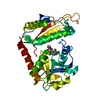
| ||||||||
|---|---|---|---|---|---|---|---|---|---|
| 1 | 
| ||||||||
| Unit cell |
|
-Components
| #1: Protein | Mass: 32321.799 Da / Num. of mol.: 1 Source method: isolated from a genetically manipulated source Source: (gene. exp.) Haliscomenobacter hydrossis DSM 1100 (bacteria)Gene: Halhy_2446 / Production host: |
|---|---|
| #2: Chemical | ChemComp-OCF /   Mass: 334.219 Da / Num. of mol.: 1 / Source method: obtained synthetically / Formula: C11H15N2O8P / Feature type: SUBJECT OF INVESTIGATION Mass: 334.219 Da / Num. of mol.: 1 / Source method: obtained synthetically / Formula: C11H15N2O8P / Feature type: SUBJECT OF INVESTIGATION |
| #3: Chemical | ChemComp-MG /   Mass: 24.305 Da / Num. of mol.: 1 / Source method: obtained synthetically / Formula: Mg Mass: 24.305 Da / Num. of mol.: 1 / Source method: obtained synthetically / Formula: Mg |
| #4: Water | ChemComp-HOH /  Mass: 18.015 Da / Num. of mol.: 307 / Source method: isolated from a natural source / Formula: H2O Mass: 18.015 Da / Num. of mol.: 307 / Source method: isolated from a natural source / Formula: H2O |
| Has ligand of interest | Y |
| Has protein modification | N |
-Experimental details
-Experiment
| Experiment | Method: X-RAY DIFFRACTION / Number of used crystals: 1 |
|---|
- Sample preparation
| Crystal | Density Matthews: 2.46 Å3/Da / Density % sol: 50.08 % |
|---|---|
| Crystal grow | Temperature: 288 K / Method: vapor diffusion, hanging drop Details: 0.2M MgCl2, 0.1M Bis-tris pH 5.5, 25% PEG 3350, 6mM PLP, 12mM 3-aminooxypropionic acid |
-Data collection
| Diffraction | Mean temperature: 100 K / Serial crystal experiment: N |
|---|---|
| Diffraction source | Source: SYNCHROTRON / Site: KURCHATOV SNC  / Beamline: K4.4 / Wavelength: 0.74503 Å / Beamline: K4.4 / Wavelength: 0.74503 Å |
| Detector | Type: MAR CCD 165 mm / Detector: CCD / Date: Jun 3, 2022 |
| Radiation | Protocol: SINGLE WAVELENGTH / Monochromatic (M) / Laue (L): M / Scattering type: x-ray |
| Radiation wavelength | Wavelength: 0.74503 Å / Relative weight: 1 |
| Reflection | Resolution: 1.7→35.34 Å / Num. obs: 32789 / % possible obs: 94.9 % / Redundancy: 3.3 % / CC1/2: 0.991 / Rmerge(I) obs: 0.083 / Rpim(I) all: 0.054 / Rrim(I) all: 0.1 / Χ2: 1.01 / Net I/σ(I): 11.4 / Num. measured all: 107399 |
| Reflection shell | Resolution: 1.7→1.73 Å / % possible obs: 98.6 % / Redundancy: 3.4 % / Rmerge(I) obs: 0.484 / Num. measured all: 6072 / Num. unique obs: 1795 / CC1/2: 0.602 / Rpim(I) all: 0.312 / Rrim(I) all: 0.578 / Χ2: 0.52 / Net I/σ(I) obs: 1.9 |
- Processing
| Software |
| ||||||||||||||||||||||||||||||||||||||||||||||||||||||||||||||||||||||||||||||||||||||||||||||||||||||||||||||||||||||||||||||||||||||||||||||||||||||||||||||||||||||||||||||||||||||
|---|---|---|---|---|---|---|---|---|---|---|---|---|---|---|---|---|---|---|---|---|---|---|---|---|---|---|---|---|---|---|---|---|---|---|---|---|---|---|---|---|---|---|---|---|---|---|---|---|---|---|---|---|---|---|---|---|---|---|---|---|---|---|---|---|---|---|---|---|---|---|---|---|---|---|---|---|---|---|---|---|---|---|---|---|---|---|---|---|---|---|---|---|---|---|---|---|---|---|---|---|---|---|---|---|---|---|---|---|---|---|---|---|---|---|---|---|---|---|---|---|---|---|---|---|---|---|---|---|---|---|---|---|---|---|---|---|---|---|---|---|---|---|---|---|---|---|---|---|---|---|---|---|---|---|---|---|---|---|---|---|---|---|---|---|---|---|---|---|---|---|---|---|---|---|---|---|---|---|---|---|---|---|---|
| Refinement | Method to determine structure: MOLECULAR REPLACEMENT / Resolution: 1.7→35.34 Å / Cor.coef. Fo:Fc: 0.963 / Cor.coef. Fo:Fc free: 0.941 / SU B: 2.51 / SU ML: 0.079 / Cross valid method: THROUGHOUT / ESU R: 0.104 / ESU R Free: 0.107 / Stereochemistry target values: MAXIMUM LIKELIHOOD / Details: HYDROGENS HAVE BEEN ADDED IN THE RIDING POSITIONS
| ||||||||||||||||||||||||||||||||||||||||||||||||||||||||||||||||||||||||||||||||||||||||||||||||||||||||||||||||||||||||||||||||||||||||||||||||||||||||||||||||||||||||||||||||||||||
| Solvent computation | Ion probe radii: 0.8 Å / Shrinkage radii: 0.8 Å / VDW probe radii: 1.2 Å / Solvent model: MASK | ||||||||||||||||||||||||||||||||||||||||||||||||||||||||||||||||||||||||||||||||||||||||||||||||||||||||||||||||||||||||||||||||||||||||||||||||||||||||||||||||||||||||||||||||||||||
| Displacement parameters | Biso mean: 16.275 Å2
| ||||||||||||||||||||||||||||||||||||||||||||||||||||||||||||||||||||||||||||||||||||||||||||||||||||||||||||||||||||||||||||||||||||||||||||||||||||||||||||||||||||||||||||||||||||||
| Refinement step | Cycle: 1 / Resolution: 1.7→35.34 Å
| ||||||||||||||||||||||||||||||||||||||||||||||||||||||||||||||||||||||||||||||||||||||||||||||||||||||||||||||||||||||||||||||||||||||||||||||||||||||||||||||||||||||||||||||||||||||
| Refine LS restraints |
|