 Movie
Movie Controller


+ Open data

- Basic information
| Entry | Database: PDB / ID: 8yrc | ||||||
|---|---|---|---|---|---|---|---|
| Title | Chlorinated YabJ from Staphylococcus aureus | ||||||
 Components Components | Translation initiation inhibitor homologue | ||||||
 Keywords Keywords | STRUCTURAL PROTEIN / ribonuclease / stress-response protein | ||||||
| Function / homology |  Function and homology information Function and homology information | ||||||
| Biological species |  Staphylococcus aureus subsp. aureus Mu50 (bacteria) Staphylococcus aureus subsp. aureus Mu50 (bacteria) | ||||||
| Method |  X-RAY DIFFRACTION / SYNCHROTRON / MOLECULAR REPLACEMENT / Resolution: 1.7 Å X-RAY DIFFRACTION / SYNCHROTRON / MOLECULAR REPLACEMENT / Resolution: 1.7 Å | ||||||
 Authors Authors | Jeong, C. / Kim, H.J. | ||||||
| Funding support | 1items
| ||||||
 Citation Citation | Journal: Biochem.Biophys.Res.Commun. / Year: 2024 Title: YabJ from Staphylococcus aureus entraps chlorides within its pocket. Authors: Jeong, C. / Kim, H.J. | ||||||
| History |
|
- Structure visualization
| Structure viewer | Molecule: MolmilJmol/JSmol |
|---|
- Downloads & links
-Download
| PDBx/mmCIF format | 8yrc.cif.gz | 86.6 KB | Display | PDBx/mmCIF format |
|---|---|---|---|---|
| PDB format | pdb8yrc.ent.gz | 64.5 KB | Display | PDB format |
| PDBx/mmJSON format | 8yrc.json.gz | Tree view | PDBx/mmJSON format | |
| Others |  Other downloads Other downloads |
-Validation report
| Summary document | 8yrc_validation.pdf.gz | 1.9 MB | Display | wwPDB validaton report |
|---|---|---|---|---|
| Full document | 8yrc_full_validation.pdf.gz | 1.9 MB | Display | |
| Data in XML | 8yrc_validation.xml.gz | 15.6 KB | Display | |
| Data in CIF | 8yrc_validation.cif.gz | 21.3 KB | Display | |
| Arichive directory | https://data.pdbj.org/pub/pdb/validation_reports/yr/8yrcftp://data.pdbj.org/pub/pdb/validation_reports/yr/8yrc | HTTPS FTP |
-Related structure data
| Related structure data | 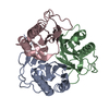 5yu2S S: Starting model for refinement |
|---|---|
| Similar structure data |
-Links
 PDBj
PDBj


- Assembly
| Deposited unit | 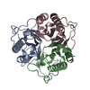
| ||||||||
|---|---|---|---|---|---|---|---|---|---|
| 1 |
| ||||||||
| Unit cell |
|
-Components
| #1: Protein | Mass: 14276.385 Da / Num. of mol.: 3 Source method: isolated from a genetically manipulated source Details: MKIINTTRLPEALGPYSHATVVNGMVYTSGQIPLNVDGKIVSADVQAQTKQVLENLKVVL EEAGSDLNSVAKATIFIKDMNDFQKINEVYGQYFNEHKPARSCVEVARLPKDVKVEIELV SKIKEL Source: (gene. exp.) Staphylococcus aureus subsp. aureus Mu50 (bacteria)Gene: SAV0497 / Production host: #2: Chemical | ChemComp-O / |   Mass: 15.999 Da / Num. of mol.: 1 / Source method: obtained synthetically / Formula: O / Feature type: SUBJECT OF INVESTIGATION Mass: 15.999 Da / Num. of mol.: 1 / Source method: obtained synthetically / Formula: O / Feature type: SUBJECT OF INVESTIGATION#3: Chemical | ChemComp-NA / |   Mass: 22.990 Da / Num. of mol.: 1 / Source method: obtained synthetically / Formula: Na / Feature type: SUBJECT OF INVESTIGATION Mass: 22.990 Da / Num. of mol.: 1 / Source method: obtained synthetically / Formula: Na / Feature type: SUBJECT OF INVESTIGATION#4: Chemical | ChemComp-CL / |   Mass: 35.453 Da / Num. of mol.: 1 / Source method: obtained synthetically / Formula: Cl / Feature type: SUBJECT OF INVESTIGATION Mass: 35.453 Da / Num. of mol.: 1 / Source method: obtained synthetically / Formula: Cl / Feature type: SUBJECT OF INVESTIGATION#5: Water | ChemComp-HOH / |  Mass: 18.015 Da / Num. of mol.: 84 / Source method: isolated from a natural source / Formula: H2O Mass: 18.015 Da / Num. of mol.: 84 / Source method: isolated from a natural source / Formula: H2OHas ligand of interest | Y | |
|---|
-Experimental details
-Experiment
| Experiment | Method: X-RAY DIFFRACTION / Number of used crystals: 1 |
|---|
- Sample preparation
| Crystal | Density Matthews: 2.34 Å3/Da / Density % sol: 47.44 % |
|---|---|
| Crystal grow | Temperature: 293 K / Method: vapor diffusion, hanging drop / Details: 25% (w/v) PEG3350 and 100 mM Tris/HCl, pH 8.5 |
-Data collection
| Diffraction | Mean temperature: 100 K / Serial crystal experiment: N |
|---|---|
| Diffraction source | Source: SYNCHROTRON / Site: PAL/PLS  / Beamline: 5C (4A) / Wavelength: 0.9795 Å / Beamline: 5C (4A) / Wavelength: 0.9795 Å |
| Detector | Type: DECTRIS EIGER X 9M / Detector: PIXEL / Date: May 4, 2023 |
| Radiation | Protocol: SINGLE WAVELENGTH / Monochromatic (M) / Laue (L): M / Scattering type: x-ray |
| Radiation wavelength | Wavelength: 0.9795 Å / Relative weight: 1 |
| Reflection | Resolution: 1.7→35.95 Å / Num. obs: 46373 / % possible obs: 97.6 % / Redundancy: 1.8 % / CC1/2: 0.99 / Net I/σ(I): 10.3 |
| Reflection shell | Resolution: 1.7→1.73 Å / Num. unique obs: 2847 / CC1/2: 0.36 |
- Processing
| Software |
| ||||||||||||||||||||||||||||||||||||||||||||||||||||||||||||||||||||||||||||||||||||||||||||||||||||||||||||||||||||||||||||||||||||||||||||||||||||||||||||||||||||||||||||||||||||||
|---|---|---|---|---|---|---|---|---|---|---|---|---|---|---|---|---|---|---|---|---|---|---|---|---|---|---|---|---|---|---|---|---|---|---|---|---|---|---|---|---|---|---|---|---|---|---|---|---|---|---|---|---|---|---|---|---|---|---|---|---|---|---|---|---|---|---|---|---|---|---|---|---|---|---|---|---|---|---|---|---|---|---|---|---|---|---|---|---|---|---|---|---|---|---|---|---|---|---|---|---|---|---|---|---|---|---|---|---|---|---|---|---|---|---|---|---|---|---|---|---|---|---|---|---|---|---|---|---|---|---|---|---|---|---|---|---|---|---|---|---|---|---|---|---|---|---|---|---|---|---|---|---|---|---|---|---|---|---|---|---|---|---|---|---|---|---|---|---|---|---|---|---|---|---|---|---|---|---|---|---|---|---|---|
| Refinement | Method to determine structure: MOLECULAR REPLACEMENT Starting model: 5YU2 Resolution: 1.7→32.51 Å / Cor.coef. Fo:Fc: 0.973 / Cor.coef. Fo:Fc free: 0.967 / SU B: 1.896 / SU ML: 0.065 / Cross valid method: THROUGHOUT / ESU R: 0.026 / ESU R Free: 0.025 / Stereochemistry target values: MAXIMUM LIKELIHOOD / Details: HYDROGENS HAVE BEEN ADDED IN THE RIDING POSITIONS
| ||||||||||||||||||||||||||||||||||||||||||||||||||||||||||||||||||||||||||||||||||||||||||||||||||||||||||||||||||||||||||||||||||||||||||||||||||||||||||||||||||||||||||||||||||||||
| Solvent computation | Ion probe radii: 0.8 Å / Shrinkage radii: 0.8 Å / VDW probe radii: 1.2 Å / Solvent model: MASK | ||||||||||||||||||||||||||||||||||||||||||||||||||||||||||||||||||||||||||||||||||||||||||||||||||||||||||||||||||||||||||||||||||||||||||||||||||||||||||||||||||||||||||||||||||||||
| Displacement parameters | Biso mean: 37.531 Å2
| ||||||||||||||||||||||||||||||||||||||||||||||||||||||||||||||||||||||||||||||||||||||||||||||||||||||||||||||||||||||||||||||||||||||||||||||||||||||||||||||||||||||||||||||||||||||
| Refinement step | Cycle: 1 / Resolution: 1.7→32.51 Å
| ||||||||||||||||||||||||||||||||||||||||||||||||||||||||||||||||||||||||||||||||||||||||||||||||||||||||||||||||||||||||||||||||||||||||||||||||||||||||||||||||||||||||||||||||||||||
| Refine LS restraints |
|