 Movie
Movie Controller

[English] 日本語
 Yorodumi
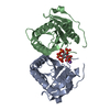
- PDB-8yjc: Structure of Vibrio vulnificus MARTX cysteine protease domain C3727A -
+ Open data

- Basic information
| Entry | Database: PDB / ID: 8yjc | ||||||
|---|---|---|---|---|---|---|---|
| Title | Structure of Vibrio vulnificus MARTX cysteine protease domain C3727A | ||||||
 Components Components | Multifunctional autoprocessing repeat-in-toxin (MARTX) | ||||||
 Keywords Keywords | TOXIN / protease / Inositol hexaphosphate / activation | ||||||
| Function / homology | INOSITOL HEXAKISPHOSPHATE Function and homology information Function and homology information | ||||||
| Biological species |  Vibrio vulnificus MO6-24/O (bacteria) Vibrio vulnificus MO6-24/O (bacteria) | ||||||
| Method |  X-RAY DIFFRACTION / SYNCHROTRON / MOLECULAR REPLACEMENT / Resolution: 1.3 Å X-RAY DIFFRACTION / SYNCHROTRON / MOLECULAR REPLACEMENT / Resolution: 1.3 Å | ||||||
 Authors Authors | Chen, L. / Khan, H. / Tan, L. / Li, X. / Zhang, G. / Im, Y.J. | ||||||
| Funding support |  Korea, Republic Of, 1items Korea, Republic Of, 1items
| ||||||
 Citation Citation | Journal: Plos One / Year: 2024 Title: Structural basis of the activation of MARTX cysteine protease domain from Vibrio vulnificus. Authors: Chen, L. / Khan, H. / Tan, L. / Li, X. / Zhang, G. / Im, Y.J. | ||||||
| History |
|
- Structure visualization
| Structure viewer | Molecule: MolmilJmol/JSmol |
|---|
- Downloads & links
-Download
| PDBx/mmCIF format | 8yjc.cif.gz | 78.7 KB | Display | PDBx/mmCIF format |
|---|---|---|---|---|
| PDB format | pdb8yjc.ent.gz | 45.6 KB | Display | PDB format |
| PDBx/mmJSON format | 8yjc.json.gz | Tree view | PDBx/mmJSON format | |
| Others |  Other downloads Other downloads |
-Validation report
| Summary document | 8yjc_validation.pdf.gz | 1014.8 KB | Display | wwPDB validaton report |
|---|---|---|---|---|
| Full document | 8yjc_full_validation.pdf.gz | 1016.5 KB | Display | |
| Data in XML | 8yjc_validation.xml.gz | 14 KB | Display | |
| Data in CIF | 8yjc_validation.cif.gz | 21.5 KB | Display | |
| Arichive directory | https://data.pdbj.org/pub/pdb/validation_reports/yj/8yjcftp://data.pdbj.org/pub/pdb/validation_reports/yj/8yjc | HTTPS FTP |
-Related structure data
-Links
 PDBj
PDBj


- Assembly
| Deposited unit | 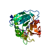
| |||||||||||||||
|---|---|---|---|---|---|---|---|---|---|---|---|---|---|---|---|---|
| 1 |
| |||||||||||||||
| Unit cell |
| |||||||||||||||
| Components on special symmetry positions |
|
-Components
| #1: Protein | Mass: 24348.615 Da / Num. of mol.: 1 / Fragment: cysteine protease domain / Mutation: C3727A Source method: isolated from a genetically manipulated source Details: NCBI Reference Sequence: WP_058645630.1, MARTX cysteine protease domain, C3727A mutant. The N-terminal GSAMGS is a linker sequence to the cleavable His-tag by thrombin. Source: (gene. exp.) Vibrio vulnificus MO6-24/O (bacteria) / Gene: rtxa / Plasmid: pHIS2-thrDetails (production host): the N-terminal thrombin cleavable His-tag fusion Production host: |
|---|---|
| #2: Chemical | ChemComp-NA /   Mass: 22.990 Da / Num. of mol.: 1 / Source method: obtained synthetically / Formula: Na Mass: 22.990 Da / Num. of mol.: 1 / Source method: obtained synthetically / Formula: Na |
| #3: Chemical | ChemComp-IHP /   Mass: 660.035 Da / Num. of mol.: 1 / Source method: obtained synthetically / Formula: C6H18O24P6 / Feature type: SUBJECT OF INVESTIGATION Mass: 660.035 Da / Num. of mol.: 1 / Source method: obtained synthetically / Formula: C6H18O24P6 / Feature type: SUBJECT OF INVESTIGATION |
| #4: Chemical | ChemComp-TRS /   Mass: 122.143 Da / Num. of mol.: 1 / Source method: obtained synthetically / Formula: C4H12NO3 / Comment: pH buffer*YM Mass: 122.143 Da / Num. of mol.: 1 / Source method: obtained synthetically / Formula: C4H12NO3 / Comment: pH buffer*YM |
| #5: Water | ChemComp-HOH /  Mass: 18.015 Da / Num. of mol.: 345 / Source method: isolated from a natural source / Formula: H2O Mass: 18.015 Da / Num. of mol.: 345 / Source method: isolated from a natural source / Formula: H2O |
| Has ligand of interest | Y |
-Experimental details
-Experiment
| Experiment | Method: X-RAY DIFFRACTION / Number of used crystals: 1 |
|---|
- Sample preparation
| Crystal | Density Matthews: 2.41 Å3/Da / Density % sol: 48.92 % |
|---|---|
| Crystal grow | Temperature: 298 K / Method: vapor diffusion, hanging drop / pH: 9 / Details: 0.1 M HEPES pH 9.0, 30% PEG 8000 |
-Data collection
| Diffraction | Mean temperature: 100 K / Serial crystal experiment: N |
|---|---|
| Diffraction source | Source: SYNCHROTRON / Site: PAL/PLS / Beamline: 7A (6B, 6C1) / Wavelength: 0.9795 Å |
| Detector | Type: ADSC QUANTUM 270 / Detector: CCD / Date: Aug 4, 2020 |
| Radiation | Protocol: SINGLE WAVELENGTH / Monochromatic (M) / Laue (L): M / Scattering type: x-ray |
| Radiation wavelength | Wavelength: 0.9795 Å / Relative weight: 1 |
| Reflection | Resolution: 1.3→50 Å / Num. obs: 54914 / % possible obs: 97.1 % / Redundancy: 4.9 % / Biso Wilson estimate: 12.96 Å2 / Rmerge(I) obs: 0.052 / Net I/σ(I): 46.8 |
| Reflection shell | Resolution: 1.3→1.32 Å / Redundancy: 3.9 % / Rmerge(I) obs: 0.263 / Mean I/σ(I) obs: 7.5 / Num. unique obs: 2163 / % possible all: 76.4 |
- Processing
| Software |
| |||||||||||||||||||||||||||||||||||||||||||||||||||||||||||||||||||||||||||||||||||||||||||||||||||||||||
|---|---|---|---|---|---|---|---|---|---|---|---|---|---|---|---|---|---|---|---|---|---|---|---|---|---|---|---|---|---|---|---|---|---|---|---|---|---|---|---|---|---|---|---|---|---|---|---|---|---|---|---|---|---|---|---|---|---|---|---|---|---|---|---|---|---|---|---|---|---|---|---|---|---|---|---|---|---|---|---|---|---|---|---|---|---|---|---|---|---|---|---|---|---|---|---|---|---|---|---|---|---|---|---|---|---|---|
| Refinement | Method to determine structure: MOLECULAR REPLACEMENT / Resolution: 1.3→24.73 Å / SU ML: 0.1106 / Cross valid method: FREE R-VALUE / σ(F): 1.35 / Phase error: 17.8738 Stereochemistry target values: GeoStd + Monomer Library + CDL v1.2
| |||||||||||||||||||||||||||||||||||||||||||||||||||||||||||||||||||||||||||||||||||||||||||||||||||||||||
| Solvent computation | Shrinkage radii: 0.9 Å / VDW probe radii: 1.11 Å / Solvent model: FLAT BULK SOLVENT MODEL | |||||||||||||||||||||||||||||||||||||||||||||||||||||||||||||||||||||||||||||||||||||||||||||||||||||||||
| Displacement parameters | Biso mean: 18.03 Å2 | |||||||||||||||||||||||||||||||||||||||||||||||||||||||||||||||||||||||||||||||||||||||||||||||||||||||||
| Refinement step | Cycle: LAST / Resolution: 1.3→24.73 Å
| |||||||||||||||||||||||||||||||||||||||||||||||||||||||||||||||||||||||||||||||||||||||||||||||||||||||||
| Refine LS restraints |
| |||||||||||||||||||||||||||||||||||||||||||||||||||||||||||||||||||||||||||||||||||||||||||||||||||||||||
| LS refinement shell |
|
