 Movie
Movie Controller

[English] 日本語
 Yorodumi
- PDB-8yif: Crystal structure of GH13_30 alpha-glucosidase CmmB in complex wi... -
+ Open data

- Basic information
| Entry | Database: PDB / ID: 8yif | ||||||
|---|---|---|---|---|---|---|---|
| Title | Crystal structure of GH13_30 alpha-glucosidase CmmB in complex with acarviosin | ||||||
 Components Components | Alpha-glucosidase | ||||||
 Keywords Keywords | HYDROLASE / alpha-glucosidase / inhibitor / complex | ||||||
| Function / homology | Oligo-1,6-glucosidase, domain 2 / alpha-amylase activity / oligosaccharide catabolic process / Alpha amylase, catalytic domain / Glycosyl hydrolase, family 13, catalytic domain / Alpha-amylase domain / Glycoside hydrolase superfamily / : / Alpha-glucosidase Function and homology information Function and homology information | ||||||
| Biological species |  Arthrobacter globiformis (bacteria) Arthrobacter globiformis (bacteria) | ||||||
| Method |  X-RAY DIFFRACTION / SYNCHROTRON / MOLECULAR REPLACEMENT / Resolution: 1.6 Å X-RAY DIFFRACTION / SYNCHROTRON / MOLECULAR REPLACEMENT / Resolution: 1.6 Å | ||||||
 Authors Authors | Saburi, W. / Tagami, T. / Yu, J. / Ose, T. / Yao, M. / Mori, H. | ||||||
| Funding support | 1items
| ||||||
 Citation Citation | Journal: Food Biosci / Year: 2024 Title: Molecular mechanism for the substrate specificity of Arthrobacter globiformis M6 alpha-glucosidase CmmB, belonging to glycoside hydrolase family 13 subfamily 30 Authors: Saburi, W. / Tagami, T. / Usui, T. / Yu, J. / Ose, T. / Yao, M. / Mori, H. | ||||||
| History |
|
- Structure visualization
| Structure viewer | Molecule: MolmilJmol/JSmol |
|---|
- Downloads & links
-Download
| PDBx/mmCIF format | 8yif.cif.gz | 130.3 KB | Display | PDBx/mmCIF format |
|---|---|---|---|---|
| PDB format | pdb8yif.ent.gz | Display | PDB format | |
| PDBx/mmJSON format | 8yif.json.gz | Tree view | PDBx/mmJSON format | |
| Others |  Other downloads Other downloads |
-Validation report
| Arichive directory | https://data.pdbj.org/pub/pdb/validation_reports/yi/8yifftp://data.pdbj.org/pub/pdb/validation_reports/yi/8yif | HTTPS FTP |
|---|
-Related structure data
| Related structure data | 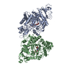 8yieSC S: Starting model for refinement C: citing same article ( |
|---|---|
| Similar structure data |
-Links
 PDBj
PDBj

- Assembly
| Deposited unit | 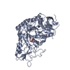
| ||||||||
|---|---|---|---|---|---|---|---|---|---|
| 1 |
| ||||||||
| Unit cell |
|
-Components
| #1: Protein | Mass: 64162.379 Da / Num. of mol.: 1 Source method: isolated from a genetically manipulated source Details: BAI67603.1 / Source: (gene. exp.) Arthrobacter globiformis (bacteria) / Gene: cmmB / Production host: |
|---|---|
| #2: Chemical | ChemComp-A1L2I / Mass: 335.350 Da / Num. of mol.: 1 Source method: isolated from a genetically manipulated source Formula: C14H25NO8 / Feature type: SUBJECT OF INVESTIGATION |
| #3: Water | ChemComp-HOH /  Mass: 18.015 Da / Num. of mol.: 425 / Source method: isolated from a natural source / Formula: H2O Mass: 18.015 Da / Num. of mol.: 425 / Source method: isolated from a natural source / Formula: H2O |
| Has ligand of interest | Y |
-Experimental details
-Experiment
| Experiment | Method: X-RAY DIFFRACTION / Number of used crystals: 1 |
|---|
- Sample preparation
| Crystal | Density Matthews: 2.14 Å3/Da / Density % sol: 42.61 % |
|---|---|
| Crystal grow | Temperature: 293 K / Method: vapor diffusion, sitting drop / pH: 7 Details: 5 mM acarviosin, 0.5 M lithium chloride, 10% (w/v) polyethylene glycol 6000, and 55 mM HEPES-NaOH buffer (pH 7.0) |
-Data collection
| Diffraction | Mean temperature: 100 K / Serial crystal experiment: N |
|---|---|
| Diffraction source | Source: SYNCHROTRON / Site: Photon Factory  / Beamline: BL-5A / Wavelength: 1 Å / Beamline: BL-5A / Wavelength: 1 Å |
| Detector | Type: DECTRIS PILATUS3 S 2M / Detector: PIXEL / Date: Mar 9, 2023 |
| Radiation | Protocol: SINGLE WAVELENGTH / Monochromatic (M) / Laue (L): M / Scattering type: x-ray |
| Radiation wavelength | Wavelength: 1 Å / Relative weight: 1 |
| Reflection | Resolution: 1.6→50 Å / Num. obs: 70932 / % possible obs: 99.6 % / Redundancy: 6.61 % / CC1/2: 0.999 / Rmerge(I) obs: 0.082 / Rrim(I) all: 0.089 / Net I/σ(I): 15.7 |
| Reflection shell | Resolution: 1.6→1.7 Å / Rmerge(I) obs: 0.672 / Mean I/σ(I) obs: 2.78 / Num. unique obs: 11229 / CC1/2: 0.873 / Rrim(I) all: 0.731 |
- Processing
| Software |
| ||||||||||||||||||||||||||||||||||||||||||||||||||||||||||||||||||||||||||||||||||||||||||||||||||
|---|---|---|---|---|---|---|---|---|---|---|---|---|---|---|---|---|---|---|---|---|---|---|---|---|---|---|---|---|---|---|---|---|---|---|---|---|---|---|---|---|---|---|---|---|---|---|---|---|---|---|---|---|---|---|---|---|---|---|---|---|---|---|---|---|---|---|---|---|---|---|---|---|---|---|---|---|---|---|---|---|---|---|---|---|---|---|---|---|---|---|---|---|---|---|---|---|---|---|---|
| Refinement | Method to determine structure: MOLECULAR REPLACEMENT Starting model: 8YIE Resolution: 1.6→42.13 Å / SU ML: 0.17 / Cross valid method: FREE R-VALUE / σ(F): 1.38 / Phase error: 19.93 / Stereochemistry target values: ML
| ||||||||||||||||||||||||||||||||||||||||||||||||||||||||||||||||||||||||||||||||||||||||||||||||||
| Solvent computation | Shrinkage radii: 0.9 Å / VDW probe radii: 1.1 Å / Solvent model: FLAT BULK SOLVENT MODEL | ||||||||||||||||||||||||||||||||||||||||||||||||||||||||||||||||||||||||||||||||||||||||||||||||||
| Refinement step | Cycle: LAST / Resolution: 1.6→42.13 Å
| ||||||||||||||||||||||||||||||||||||||||||||||||||||||||||||||||||||||||||||||||||||||||||||||||||
| Refine LS restraints |
| ||||||||||||||||||||||||||||||||||||||||||||||||||||||||||||||||||||||||||||||||||||||||||||||||||
| LS refinement shell |
|
