[English] 日本語
Yorodumi
- PDB-8ygw: The Crystal Structure of MAPK11 from Biortus -

+
Open data


ID or keywords:

Loading...

-
Basic information

Entry
Database: PDB / ID: 8ygw
TitleThe Crystal Structure of MAPK11 from Biortus
ComponentsMitogen-activated protein kinase 11
KeywordsTRANSCRIPTION / Kinase / Serine/threonine-protein kinase / Transferase / Stress response / Transcription regulation / ATP-binding / Nucleotide-binding
Function / homology
Function and homology information


negative regulation of cardiac muscle cell proliferation / stress-activated protein kinase signaling cascade / Activation of PPARGC1A (PGC-1alpha) by phosphorylation / cardiac muscle cell proliferation / KSRP (KHSRP) binds and destabilizes mRNA / DSCAM interactions / cellular response to UV-B / positive regulation of muscle cell differentiation / Myogenesis / Activation of the AP-1 family of transcription factors ...negative regulation of cardiac muscle cell proliferation / stress-activated protein kinase signaling cascade / Activation of PPARGC1A (PGC-1alpha) by phosphorylation / cardiac muscle cell proliferation / KSRP (KHSRP) binds and destabilizes mRNA / DSCAM interactions / cellular response to UV-B / positive regulation of muscle cell differentiation / Myogenesis / Activation of the AP-1 family of transcription factors / ERK/MAPK targets / p38MAPK cascade / RHO GTPases Activate NADPH Oxidases / MAP kinase activity / mitogen-activated protein kinase / cellular response to interleukin-1 / stress-activated MAPK cascade / p38MAPK events / positive regulation of interleukin-12 production / activated TAK1 mediates p38 MAPK activation / NOD1/2 Signaling Pathway / bone development / cellular response to virus / VEGFA-VEGFR2 Pathway / osteoblast differentiation / cellular senescence / Oxidative Stress Induced Senescence / Regulation of TP53 Activity through Phosphorylation / intracellular signal transduction / phosphorylation / protein serine kinase activity / protein serine/threonine kinase activity / positive regulation of gene expression / nucleoplasm / ATP binding / nucleus / cytoplasm / cytosol
Similarity search - Function
Mitogen-activated protein (MAP) kinase p38-like / Mitogen-activated protein (MAP) kinase, conserved site / MAP kinase signature. / Protein kinase domain / Serine/Threonine protein kinases, catalytic domain / Protein kinase, ATP binding site / Protein kinases ATP-binding region signature. / Protein kinase domain profile. / Protein kinase domain / Protein kinase-like domain superfamily
Similarity search - Domain/homology
Chem-B96 / Mitogen-activated protein kinase 11
Similarity search - Component
Biological speciesHomo sapiens (human)
MethodX-RAY DIFFRACTION / SYNCHROTRON / MOLECULAR REPLACEMENT / Resolution: 3.301 Å
AuthorsWang, F. / Cheng, W. / Yuan, Z. / Lin, D. / Guo, S.
Funding support1items
OrganizationGrant numberCountry
Not funded
CitationJournal: To Be Published
Title: The Crystal Structure of MAPK11 from Biortus.
Authors: Wang, F. / Cheng, W. / Yuan, Z. / Lin, D. / Guo, S.
History
DepositionFeb 27, 2024Deposition site: PDBJ / Processing site: PDBC
Revision 1.0Mar 13, 2024Provider: repository / Type: Initial release

-
Structure visualization

Structure viewerMolecule:
MolmilJmol/JSmol

Downloads & links

-
Assembly

Deposited unit
A: Mitogen-activated protein kinase 11
hetero molecules


Theoretical massNumber of molelcules
Total (without water)41,9352
Polymers41,4071
Non-polymers5281
Water46826
1


  • Idetical with deposited unit
  • defined by author&software
  • Evidence: gel filtration
TypeNameSymmetry operationNumber
identity operation1_555x,y,z1
Buried area1210 Å2
ΔGint-6 kcal/mol
Surface area15940 Å2
MethodPISA
Unit cell
Length a, b, c (Å)39.061, 61.860, 147.437
Angle α, β, γ (deg.)90.000, 90.000, 90.000
Int Tables number19
Space group name H-MP212121

-
Components

#1: Protein Mitogen-activated protein kinase 11 / MAP kinase 11 / MAPK 11 / Mitogen-activated protein kinase p38 beta / MAP kinase p38 beta / p38b / ...MAP kinase 11 / MAPK 11 / Mitogen-activated protein kinase p38 beta / MAP kinase p38 beta / p38b / Stress-activated protein kinase 2b / SAPK2b / p38-2


Mass: 41407.168 Da / Num. of mol.: 1
Source method: isolated from a genetically manipulated source
Source: (gene. exp.) Homo sapiens (human) / Gene: MAPK11, PRKM11, SAPK2, SAPK2B / Production host: Escherichia coli (E. coli)
References: UniProt: Q15759, mitogen-activated protein kinase
#2: Chemical ChemComp-B96 / 1-(5-TERT-BUTYL-2-P-TOLYL-2H-PYRAZOL-3-YL)-3-[4-(2-MORPHOLIN-4-YL-ETHOXY)-NAPHTHALEN-1-YL]-UREA


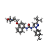
Mass: 527.657 Da / Num. of mol.: 1 / Source method: obtained synthetically / Formula: C31H37N5O3 / Feature type: SUBJECT OF INVESTIGATION
#3: Water ChemComp-HOH / water


Mass: 18.015 Da / Num. of mol.: 26 / Source method: isolated from a natural source / Formula: H2O
Has ligand of interestY

-
Experimental details

-
Experiment

ExperimentMethod: X-RAY DIFFRACTION / Number of used crystals: 1

-
Sample preparation

CrystalDensity Matthews: 2.15 Å3/Da / Density % sol: 42.82 %
Crystal growTemperature: 293 K / Method: vapor diffusion, sitting drop / Details: 0.1M Bis-Tris pH6.5, 28% PEGMME2,000

-
Data collection

DiffractionMean temperature: 100 K / Serial crystal experiment: N
Diffraction sourceSource: SYNCHROTRON / Site: CLSI / Beamline: 08ID-1 / Wavelength: 0.95374 Å
DetectorType: DECTRIS PILATUS3 6M / Detector: PIXEL / Date: May 27, 2023
RadiationProtocol: SINGLE WAVELENGTH / Monochromatic (M) / Laue (L): M / Scattering type: x-ray
Radiation wavelengthWavelength: 0.95374 Å / Relative weight: 1
ReflectionResolution: 3.3→49.15 Å / Num. obs: 5815 / % possible obs: 100 % / Redundancy: 7.3 % / Rmerge(I) obs: 0.199 / Net I/σ(I): 9.3
Reflection shellResolution: 3.3→3.57 Å / Rmerge(I) obs: 0.827 / Num. unique obs: 1159

-
Processing

Software
NameVersionClassification
REFMAC5.8.0352refinement
XDSdata reduction
Aimlessdata scaling
PHASERphasing
RefinementMethod to determine structure: MOLECULAR REPLACEMENT / Resolution: 3.301→49.15 Å / Cor.coef. Fo:Fc: 0.936 / Cor.coef. Fo:Fc free: 0.878 / SU B: 35.3 / SU ML: 0.538 / Cross valid method: FREE R-VALUE / ESU R Free: 0.654
Details: Hydrogens have been added in their riding positions
RfactorNum. reflection% reflection
Rfree0.2781 284 4.913 %
Rwork0.2054 5496 -
all0.209 --
obs-5780 99.948 %
Solvent computationIon probe radii: 0.8 Å / Shrinkage radii: 0.8 Å / VDW probe radii: 1.2 Å / Solvent model: MASK BULK SOLVENT
Displacement parametersBiso mean: 78.808 Å2
Baniso -1Baniso -2Baniso -3
1--2.978 Å20 Å20 Å2
2--7.254 Å2-0 Å2
3----4.276 Å2
Refinement stepCycle: LAST / Resolution: 3.301→49.15 Å
ProteinNucleic acidLigandSolventTotal
Num. atoms2632 0 39 26 2697
Refine LS restraints
Refine-IDTypeDev idealDev ideal targetNumber
X-RAY DIFFRACTIONr_bond_refined_d0.0020.0122732
X-RAY DIFFRACTIONr_bond_other_d0.0010.0162513
X-RAY DIFFRACTIONr_angle_refined_deg0.6851.6493705
X-RAY DIFFRACTIONr_angle_other_deg0.2391.5625850
X-RAY DIFFRACTIONr_dihedral_angle_1_deg5.2815323
X-RAY DIFFRACTIONr_dihedral_angle_2_deg2.609522
X-RAY DIFFRACTIONr_dihedral_angle_3_deg11.10710473
X-RAY DIFFRACTIONr_dihedral_angle_6_deg10.80110130
X-RAY DIFFRACTIONr_chiral_restr0.0320.2407
X-RAY DIFFRACTIONr_gen_planes_refined0.0020.023081
X-RAY DIFFRACTIONr_gen_planes_other0.0010.02542
X-RAY DIFFRACTIONr_nbd_refined0.1790.2598
X-RAY DIFFRACTIONr_symmetry_nbd_other0.1690.22597
X-RAY DIFFRACTIONr_nbtor_refined0.1680.21363
X-RAY DIFFRACTIONr_symmetry_nbtor_other0.0720.21444
X-RAY DIFFRACTIONr_xyhbond_nbd_refined0.1320.258
X-RAY DIFFRACTIONr_symmetry_nbd_refined0.120.211
X-RAY DIFFRACTIONr_nbd_other0.1570.238
X-RAY DIFFRACTIONr_symmetry_xyhbond_nbd_refined0.1130.24
X-RAY DIFFRACTIONr_mcbond_it2.0498.2021301
X-RAY DIFFRACTIONr_mcbond_other2.0488.2031301
X-RAY DIFFRACTIONr_mcangle_it3.60612.3021621
X-RAY DIFFRACTIONr_mcangle_other3.60612.3021622
X-RAY DIFFRACTIONr_scbond_it1.5988.261431
X-RAY DIFFRACTIONr_scbond_other1.5978.2571432
X-RAY DIFFRACTIONr_scangle_it2.86212.3332084
X-RAY DIFFRACTIONr_scangle_other2.86112.3312085
X-RAY DIFFRACTIONr_lrange_it6.091103.5643126
X-RAY DIFFRACTIONr_lrange_other6.09103.5543127
LS refinement shell
Resolution (Å)Rfactor RfreeNum. reflection RfreeRfactor RworkNum. reflection RworkRefine-ID% reflection obs (%)
3.301-3.3860.35150.277378X-RAY DIFFRACTION100
3.386-3.4780.339310.275388X-RAY DIFFRACTION100
3.478-3.5780.34390.274386X-RAY DIFFRACTION100
3.578-3.6880.291140.275361X-RAY DIFFRACTION100
3.688-3.8080.344210.241349X-RAY DIFFRACTION100
3.808-3.9410.225170.233350X-RAY DIFFRACTION99.7283
3.941-4.0890.347140.21328X-RAY DIFFRACTION100
4.089-4.2540.33170.195318X-RAY DIFFRACTION100
4.254-4.4420.294200.188311X-RAY DIFFRACTION100
4.442-4.6570.253200.17285X-RAY DIFFRACTION100
4.657-4.9060.266180.185293X-RAY DIFFRACTION100
4.906-5.20.19190.176266X-RAY DIFFRACTION100
5.2-5.5540.196120.175259X-RAY DIFFRACTION100
5.554-5.9920.273100.196228X-RAY DIFFRACTION100
5.992-6.5540.258140.208237X-RAY DIFFRACTION100
6.554-7.310.34160.163190X-RAY DIFFRACTION100
7.31-8.4080.27120.172181X-RAY DIFFRACTION100
8.408-10.2170.36650.151168X-RAY DIFFRACTION100
10.217-14.1250.13980.145128X-RAY DIFFRACTION100
14.125-49.150.30220.32892X-RAY DIFFRACTION100

+
About Yorodumi

-
News

-
Feb 9, 2022. New format data for meta-information of EMDB entries

New format data for meta-information of EMDB entries

  • Version 3 of the EMDB header file is now the official format.
  • The previous official version 1.9 will be removed from the archive.

Related info.:EMDB header

External links:wwPDB to switch to version 3 of the EMDB data model

-
Aug 12, 2020. Covid-19 info

Covid-19 info

URL: https://pdbjlvh1.pdbj.org/emnavi/covid19.php

New page: Covid-19 featured information page in EM Navigator.

Related info.:Covid-19 info / Mar 5, 2020. Novel coronavirus structure data

+
Mar 5, 2020. Novel coronavirus structure data

Novel coronavirus structure data

Related info.:Yorodumi Speices / Aug 12, 2020. Covid-19 info

External links:COVID-19 featured content - PDBj / Molecule of the Month (242):Coronavirus Proteases

+
Jan 31, 2019. EMDB accession codes are about to change! (news from PDBe EMDB page)

EMDB accession codes are about to change! (news from PDBe EMDB page)

  • The allocation of 4 digits for EMDB accession codes will soon come to an end. Whilst these codes will remain in use, new EMDB accession codes will include an additional digit and will expand incrementally as the available range of codes is exhausted. The current 4-digit format prefixed with “EMD-” (i.e. EMD-XXXX) will advance to a 5-digit format (i.e. EMD-XXXXX), and so on. It is currently estimated that the 4-digit codes will be depleted around Spring 2019, at which point the 5-digit format will come into force.
  • The EM Navigator/Yorodumi systems omit the EMD- prefix.

Related info.:Q: What is EMD? / ID/Accession-code notation in Yorodumi/EM Navigator

External links:EMDB Accession Codes are Changing Soon! / Contact to PDBj

+
Jul 12, 2017. Major update of PDB

Major update of PDB

  • wwPDB released updated PDB data conforming to the new PDBx/mmCIF dictionary.
  • This is a major update changing the version number from 4 to 5, and with Remediation, in which all the entries are updated.
  • In this update, many items about electron microscopy experimental information are reorganized (e.g. em_software).
  • Now, EM Navigator and Yorodumi are based on the updated data.

External links:wwPDB Remediation / Enriched Model Files Conforming to OneDep Data Standards Now Available in the PDB FTP Archive

-
Yorodumi

Thousand views of thousand structures

  • Yorodumi is a browser for structure data from EMDB, PDB, SASBDB, etc.
  • This page is also the successor to EM Navigator detail page, and also detail information page/front-end page for Omokage search.
  • The word "yorodu" (or yorozu) is an old Japanese word meaning "ten thousand". "mi" (miru) is to see.

Related info.:EMDB / PDB / SASBDB / Comparison of 3 databanks / Yorodumi Search / Aug 31, 2016. New EM Navigator & Yorodumi / Yorodumi Papers / Jmol/JSmol / Function and homology information / Changes in new EM Navigator and Yorodumi

Read more