 Movie
Movie Controller


+ Open data

- Basic information
| Entry | Database: PDB / ID: 8ygd | ||||||||||||||||||||||||
|---|---|---|---|---|---|---|---|---|---|---|---|---|---|---|---|---|---|---|---|---|---|---|---|---|---|
| Title | Rhodobacter blasticus RC-LH1 dimer | ||||||||||||||||||||||||
 Components Components |
| ||||||||||||||||||||||||
 Keywords Keywords | PHOTOSYNTHESIS / light-harvesting complex | ||||||||||||||||||||||||
| Function / homology |  Function and homology information Function and homology informationplasma membrane-derived chromatophore membrane / plasma membrane light-harvesting complex / bacteriochlorophyll binding / photosynthetic electron transport in photosystem II / photosynthesis, light reaction / metal ion binding / plasma membrane Similarity search - Function | ||||||||||||||||||||||||
| Biological species |  Fuscovulum blasticum DSM 2131 (bacteria) Fuscovulum blasticum DSM 2131 (bacteria) | ||||||||||||||||||||||||
| Method | ELECTRON MICROSCOPY / single particle reconstruction / cryo EM / Resolution: 2.84 Å | ||||||||||||||||||||||||
 Authors Authors | Liu, L.N. / Zhang, Y.Z. / Wang, P. / Christianson, B.M. / Ugurlar, D. | ||||||||||||||||||||||||
| Funding support |  China, 1items China, 1items
| ||||||||||||||||||||||||
 Citation Citation | Journal: Sci Adv / Year: 2024 Title: Architectures of photosynthetic RC-LH1 supercomplexes from . Authors: Peng Wang / Bern M Christianson / Deniz Ugurlar / Ruichao Mao / Yi Zhang / Ze-Kun Liu / Ying-Yue Zhang / Adrian M Gardner / Jun Gao / Yu-Zhong Zhang / Lu-Ning Liu /   Abstract: The reaction center-light-harvesting complex 1 (RC-LH1) plays an essential role in the primary reactions of bacterial photosynthesis. Here, we present high-resolution structures of native monomeric ...The reaction center-light-harvesting complex 1 (RC-LH1) plays an essential role in the primary reactions of bacterial photosynthesis. Here, we present high-resolution structures of native monomeric and dimeric RC-LH1 supercomplexes from () using cryo-electron microscopy. The RC-LH1 monomer is composed of an RC encircled by an open LH1 ring comprising 15 αβ heterodimers and a PufX transmembrane polypeptide. In the RC-LH1 dimer, two crossing PufX polypeptides mediate dimerization. Unlike counterpart, RC-LH1 dimer has a less bent conformation, lacks the PufY subunit near the LH1 opening, and includes two extra LH1 αβ subunits, forming a more enclosed S-shaped LH1 ring. Spectroscopic assays reveal that these unique structural features are accompanied by changes in the kinetics of quinone/quinol trafficking between RC-LH1 and cytochrome . Our findings reveal the assembly principles and structural variability of photosynthetic RC-LH1 supercomplexes, highlighting diverse strategies used by phototrophic bacteria to optimize light-harvesting and electron transfer in competitive environments. | ||||||||||||||||||||||||
| History |
|
- Structure visualization
| Structure viewer | Molecule: MolmilJmol/JSmol |
|---|
- Downloads & links
-Download
| PDBx/mmCIF format | 8ygd.cif.gz | 515.7 KB | Display | PDBx/mmCIF format |
|---|---|---|---|---|
| PDB format | pdb8ygd.ent.gz | 440.9 KB | Display | PDB format |
| PDBx/mmJSON format | 8ygd.json.gz | Tree view | PDBx/mmJSON format | |
| Others |  Other downloads Other downloads |
-Validation report
| Arichive directory | https://data.pdbj.org/pub/pdb/validation_reports/yg/8ygdftp://data.pdbj.org/pub/pdb/validation_reports/yg/8ygd | HTTPS FTP |
|---|
-Related structure data
| Related structure data |  39244MC 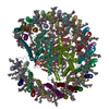 8yglC M: map data used to model this data C: citing same article ( |
|---|---|
| Similar structure data |
-Links
 PDBj
PDBj


- Assembly
| Deposited unit | 
|
|---|---|
| 1 | 
|
-Components
-Antenna pigment protein ... , 2 types, 30 molecules 028BCEGJNPRTVZb1379ADFIKOQSUWa
| #1: Protein/peptide | Mass: 5614.452 Da / Num. of mol.: 15 / Source method: isolated from a natural source / Source: (natural) Fuscovulum blasticum DSM 2131 (bacteria) / References: UniProt: A0A2T4JAH7#2: Protein | Mass: 7096.406 Da / Num. of mol.: 15 / Source method: isolated from a natural source / Source: (natural) Fuscovulum blasticum DSM 2131 (bacteria) / References: UniProt: A0A2T4JA00 |
|---|
-Protein , 2 types, 2 molecules HX
| #3: Protein | Mass: 28320.168 Da / Num. of mol.: 1 / Source method: isolated from a natural source / Source: (natural) Fuscovulum blasticum DSM 2131 (bacteria) / References: UniProt: A0A2T4J4Z7 |
|---|---|
| #6: Protein | Mass: 8046.359 Da / Num. of mol.: 1 / Source method: isolated from a natural source / Source: (natural) Fuscovulum blasticum DSM 2131 (bacteria) / References: UniProt: A0A2T4J9W4 |
-Reaction center protein ... , 2 types, 2 molecules LM
| #4: Protein | Mass: 31494.471 Da / Num. of mol.: 1 / Source method: isolated from a natural source / Source: (natural) Fuscovulum blasticum DSM 2131 (bacteria) / References: UniProt: A0A2L1K3X9 |
|---|---|
| #5: Protein | Mass: 34542.602 Da / Num. of mol.: 1 / Source method: isolated from a natural source / Source: (natural) Fuscovulum blasticum DSM 2131 (bacteria) / References: UniProt: A0A2T4J9V9 |
-Non-polymers , 7 types, 87 molecules 


| #7: Chemical | ChemComp-BCL /  Mass: 911.504 Da / Num. of mol.: 34 / Source method: obtained synthetically / Formula: C55H74MgN4O6 / Feature type: SUBJECT OF INVESTIGATION Mass: 911.504 Da / Num. of mol.: 34 / Source method: obtained synthetically / Formula: C55H74MgN4O6 / Feature type: SUBJECT OF INVESTIGATION#8: Chemical | ChemComp-SPO /  Mass: 568.914 Da / Num. of mol.: 29 / Source method: obtained synthetically / Formula: C41H60O / Feature type: SUBJECT OF INVESTIGATION Mass: 568.914 Da / Num. of mol.: 29 / Source method: obtained synthetically / Formula: C41H60O / Feature type: SUBJECT OF INVESTIGATION#9: Chemical | ChemComp-PC1 /  Mass: 790.145 Da / Num. of mol.: 11 / Source method: obtained synthetically / Formula: C44H88NO8P / Feature type: SUBJECT OF INVESTIGATION / Comment: phospholipid*YM Mass: 790.145 Da / Num. of mol.: 11 / Source method: obtained synthetically / Formula: C44H88NO8P / Feature type: SUBJECT OF INVESTIGATION / Comment: phospholipid*YM#10: Chemical |  Mass: 1464.043 Da / Num. of mol.: 3 / Source method: obtained synthetically / Formula: C81H156O17P2 / Feature type: SUBJECT OF INVESTIGATION / Comment: phospholipid*YM Mass: 1464.043 Da / Num. of mol.: 3 / Source method: obtained synthetically / Formula: C81H156O17P2 / Feature type: SUBJECT OF INVESTIGATION / Comment: phospholipid*YM#11: Chemical |  Mass: 889.215 Da / Num. of mol.: 2 / Source method: obtained synthetically / Formula: C55H76N4O6 / Feature type: SUBJECT OF INVESTIGATION Mass: 889.215 Da / Num. of mol.: 2 / Source method: obtained synthetically / Formula: C55H76N4O6 / Feature type: SUBJECT OF INVESTIGATION#12: Chemical | ChemComp-U10 /  Mass: 863.343 Da / Num. of mol.: 7 / Source method: obtained synthetically / Formula: C59H90O4 / Feature type: SUBJECT OF INVESTIGATION Mass: 863.343 Da / Num. of mol.: 7 / Source method: obtained synthetically / Formula: C59H90O4 / Feature type: SUBJECT OF INVESTIGATION#13: Chemical | ChemComp-FE2 / |  Mass: 55.845 Da / Num. of mol.: 1 / Source method: obtained synthetically / Formula: Fe / Feature type: SUBJECT OF INVESTIGATION Mass: 55.845 Da / Num. of mol.: 1 / Source method: obtained synthetically / Formula: Fe / Feature type: SUBJECT OF INVESTIGATION |
|---|
-Details
| Has ligand of interest | Y |
|---|---|
| Has protein modification | N |
-Experimental details
-Experiment
| Experiment | Method: ELECTRON MICROSCOPY |
|---|---|
| EM experiment | Aggregation state: PARTICLE / 3D reconstruction method: single particle reconstruction |
- Sample preparation
| Component | Name: Rhodobacter blasticus RC-LH1 dimer / Type: COMPLEX / Entity ID: #1-#6 / Source: NATURAL |
|---|---|
| Source (natural) | Organism: Fuscovulum blasticum DSM 2131 (bacteria) |
| Buffer solution | pH: 8 |
| Specimen | Embedding applied: NO / Shadowing applied: NO / Staining applied: NO / Vitrification applied: YES |
| Vitrification | Cryogen name: ETHANE |
- Electron microscopy imaging
| Experimental equipment |  Model: Titan Krios / Image courtesy: FEI Company |
|---|---|
| Microscopy | Model: FEI TITAN KRIOS |
| Electron gun | Electron source:  FIELD EMISSION GUN / Accelerating voltage: 300 kV / Illumination mode: FLOOD BEAM FIELD EMISSION GUN / Accelerating voltage: 300 kV / Illumination mode: FLOOD BEAM |
| Electron lens | Mode: BRIGHT FIELD / Nominal defocus max: 2000 nm / Nominal defocus min: 800 nm |
| Image recording | Electron dose: 40 e/Å2 / Film or detector model: FEI FALCON IV (4k x 4k) |
- Processing
| EM software | Name: PHENIX |
|---|---|
| CTF correction | Type: PHASE FLIPPING AND AMPLITUDE CORRECTION |
| Symmetry | Point symmetry: C2 (2 fold cyclic) |
| 3D reconstruction | Resolution: 2.84 Å / Resolution method: FSC 0.143 CUT-OFF / Num. of particles: 9149 / Symmetry type: POINT |
| Refinement | Highest resolution: 2.84 Å |
