 Movie
Movie Controller


+ Open data

- Basic information
| Entry | Database: PDB / ID: 8yfy | ||||||
|---|---|---|---|---|---|---|---|
| Title | CRYSTAL STRUCTURE OF THE EST1 H274D MUTANT AT PH 4.2 | ||||||
 Components Components | Carboxylesterase | ||||||
 Keywords Keywords | HYDROLASE / ALPHA/BETA-HYDRORASE FOLD / CARBOXYLESTERASE | ||||||
| Function / homology | Lipase, GDXG, putative serine active site / Lipolytic enzymes "G-D-X-G" family, putative serine active site. / carboxylesterase / : / Alpha/beta hydrolase fold-3 / alpha/beta hydrolase fold / carboxylesterase activity / Alpha/Beta hydrolase fold / Carboxylesterase Function and homology information Function and homology information | ||||||
| Biological species |   Saccharolobus shibatae (archaea) Saccharolobus shibatae (archaea) | ||||||
| Method |  X-RAY DIFFRACTION / SYNCHROTRON / MOLECULAR REPLACEMENT / Resolution: 1.45 Å X-RAY DIFFRACTION / SYNCHROTRON / MOLECULAR REPLACEMENT / Resolution: 1.45 Å | ||||||
 Authors Authors | Unno, H. / Oshima, Y. / Nishino, T. / Nakayama, T. / Kusunoki, M. | ||||||
| Funding support |  Japan, 1items Japan, 1items
| ||||||
 Citation Citation | Journal: J.Biosci.Bioeng. / Year: 2024 Title: Lowering pH optimum of activity of SshEstI, a slightly alkaliphilic archaeal esterase of the hormone-sensitive lipase family. Authors: Ohara, K. / Oshima, Y. / Unno, H. / Nagano, S. / Kusunoki, M. / Takahashi, S. / Waki, T. / Yamashita, S. / Nakayama, T. | ||||||
| History |
|
- Structure visualization
| Structure viewer | Molecule: MolmilJmol/JSmol |
|---|
- Downloads & links
-Download
| PDBx/mmCIF format | 8yfy.cif.gz | 79.6 KB | Display | PDBx/mmCIF format |
|---|---|---|---|---|
| PDB format | pdb8yfy.ent.gz | 56.8 KB | Display | PDB format |
| PDBx/mmJSON format | 8yfy.json.gz | Tree view | PDBx/mmJSON format | |
| Others |  Other downloads Other downloads |
-Validation report
| Arichive directory | https://data.pdbj.org/pub/pdb/validation_reports/yf/8yfyftp://data.pdbj.org/pub/pdb/validation_reports/yf/8yfy | HTTPS FTP |
|---|
-Related structure data
| Related structure data | 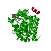 8yfzC  1wzj 1wzp 1wzq 1wzr 1wzs C: citing same article ( |
|---|---|
| Similar structure data |
-Links
 PDBj
PDBj- Assembly
| Deposited unit | 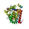
| |||||||||
|---|---|---|---|---|---|---|---|---|---|---|
| 1 | 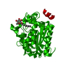
| |||||||||
| Unit cell |
| |||||||||
| Components on special symmetry positions |
|
-Components
| #1: Protein | Mass: 33528.344 Da / Num. of mol.: 1 / Mutation: H274D Source method: isolated from a genetically manipulated source Source: (gene. exp.) Saccharolobus shibatae (archaea) / Gene: SshEstI, J5U21_01394, J5U22_01308 / Plasmid: PTC99A / Production host:  |
|---|---|
| #2: Sugar | ChemComp-BOG /   Type: D-saccharide / Mass: 292.369 Da / Num. of mol.: 1 Type: D-saccharide / Mass: 292.369 Da / Num. of mol.: 1Source method: isolated from a genetically manipulated source Formula: C14H28O6 / Feature type: SUBJECT OF INVESTIGATION / Comment: detergent*YM |
| #3: Water | ChemComp-HOH /  Mass: 18.015 Da / Num. of mol.: 259 / Source method: isolated from a natural source / Formula: H2O Mass: 18.015 Da / Num. of mol.: 259 / Source method: isolated from a natural source / Formula: H2O |
| Has ligand of interest | Y |
-Experimental details
-Experiment
| Experiment | Method: X-RAY DIFFRACTION / Number of used crystals: 1 |
|---|
- Sample preparation
| Crystal | Density Matthews: 2.2 Å3/Da / Density % sol: 42.4 % |
|---|---|
| Crystal grow | Temperature: 293 K / Method: vapor diffusion / pH: 4.2 Details: 12% PEG 3000, 200 MM NACL, 100 MM PHOSPHATE-CITRATE, PH 4.2, VAPOR DIFFUSION, TEMPERATURE 293K PH range: 4.2 |
-Data collection
| Diffraction | Mean temperature: 90 K / Serial crystal experiment: N |
|---|---|
| Diffraction source | Source: SYNCHROTRON / Site: SPring-8 / Beamline: BL44XU / Wavelength: 0.9 Å |
| Detector | Type: Bruker DIP-6040 / Detector: IMAGE PLATE / Date: Sep 22, 2003 |
| Radiation | Monochromator: SI(111) / Protocol: SINGLE WAVELENGTH / Monochromatic (M) / Laue (L): M / Scattering type: x-ray |
| Radiation wavelength | Wavelength: 0.9 Å / Relative weight: 1 |
| Reflection | Resolution: 1.45→18.7 Å / Num. obs: 46275 / % possible obs: 91.5 % / Observed criterion σ(I): 0 / Redundancy: 4.3 % / Biso Wilson estimate: 18.15 Å2 / Rmerge(I) obs: 0.088 / Net I/σ(I): 13.6 |
| Reflection shell | Resolution: 1.45→1.53 Å / Redundancy: 3.6 % / Rmerge(I) obs: 0.234 / Mean I/σ(I) obs: 4.6 / Num. unique obs: 46275 / % possible all: 78.6 |
- Processing
| Software |
| ||||||||||||||||||||||||||||||||||||||||||||||||||||||||||||||||||||||||||||||||||||||||||||||||||||||||||||||||||||||||||||||||||||||||||||||||||||||||||||||||||||||||||||||||||||||
|---|---|---|---|---|---|---|---|---|---|---|---|---|---|---|---|---|---|---|---|---|---|---|---|---|---|---|---|---|---|---|---|---|---|---|---|---|---|---|---|---|---|---|---|---|---|---|---|---|---|---|---|---|---|---|---|---|---|---|---|---|---|---|---|---|---|---|---|---|---|---|---|---|---|---|---|---|---|---|---|---|---|---|---|---|---|---|---|---|---|---|---|---|---|---|---|---|---|---|---|---|---|---|---|---|---|---|---|---|---|---|---|---|---|---|---|---|---|---|---|---|---|---|---|---|---|---|---|---|---|---|---|---|---|---|---|---|---|---|---|---|---|---|---|---|---|---|---|---|---|---|---|---|---|---|---|---|---|---|---|---|---|---|---|---|---|---|---|---|---|---|---|---|---|---|---|---|---|---|---|---|---|---|---|
| Refinement | Method to determine structure: MOLECULAR REPLACEMENT / Resolution: 1.45→18.7 Å / Cor.coef. Fo:Fc: 0.973 / Cor.coef. Fo:Fc free: 0.964 / SU B: 0.935 / SU ML: 0.037 / Cross valid method: THROUGHOUT / σ(F): 0 / ESU R: 0.066 / ESU R Free: 0.067 / Stereochemistry target values: MAXIMUM LIKELIHOOD / Details: HYDROGENS HAVE BEEN ADDED IN THE RIDING POSITIONS
| ||||||||||||||||||||||||||||||||||||||||||||||||||||||||||||||||||||||||||||||||||||||||||||||||||||||||||||||||||||||||||||||||||||||||||||||||||||||||||||||||||||||||||||||||||||||
| Solvent computation | Ion probe radii: 0.8 Å / Shrinkage radii: 0.8 Å / VDW probe radii: 1.2 Å / Solvent model: BABINET MODEL WITH MASK | ||||||||||||||||||||||||||||||||||||||||||||||||||||||||||||||||||||||||||||||||||||||||||||||||||||||||||||||||||||||||||||||||||||||||||||||||||||||||||||||||||||||||||||||||||||||
| Displacement parameters | Biso mean: 16.65 Å2
| ||||||||||||||||||||||||||||||||||||||||||||||||||||||||||||||||||||||||||||||||||||||||||||||||||||||||||||||||||||||||||||||||||||||||||||||||||||||||||||||||||||||||||||||||||||||
| Refinement step | Cycle: LAST / Resolution: 1.45→18.7 Å
| ||||||||||||||||||||||||||||||||||||||||||||||||||||||||||||||||||||||||||||||||||||||||||||||||||||||||||||||||||||||||||||||||||||||||||||||||||||||||||||||||||||||||||||||||||||||
| Refine LS restraints |
|