National Natural Science Foundation of China (NSFC)
32271253
China
Citation
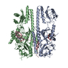
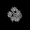
Journal: Cell / Year: 2024 Title: Light-induced remodeling of phytochrome B enables signal transduction by phytochrome-interacting factor. Authors: Zhengdong Wang / Wenfeng Wang / Didi Zhao / Yanping Song / Xiaoli Lin / Meng Shen / Cheng Chi / Bin Xu / Jun Zhao / Xing Wang Deng / Jizong Wang / Abstract: Phytochrome B (phyB) and phytochrome-interacting factors (PIFs) constitute a well-established signaling module critical for plants adapting to ambient light. However, mechanisms underlying phyB ...Phytochrome B (phyB) and phytochrome-interacting factors (PIFs) constitute a well-established signaling module critical for plants adapting to ambient light. However, mechanisms underlying phyB photoactivation and PIF binding for signal transduction remain elusive. Here, we report the cryo-electron microscopy (cryo-EM) structures of the photoactivated phyB or the constitutively active phyB mutant in complex with PIF6, revealing a similar trimer. The light-induced configuration switch of the chromophore drives a conformational transition of the nearby tongue signature within the phytochrome-specific (PHY) domain of phyB. The resulting α-helical PHY tongue further disrupts the head-to-tail dimer of phyB in the dark-adapted state. These structural remodelings of phyB facilitate the induced-fit recognition of PIF6, consequently stabilizing the N-terminal extension domain and a head-to-head dimer of activated phyB. Interestingly, the phyB dimer exhibits slight asymmetry, resulting in the binding of only one PIF6 molecule. Overall, our findings solve a key question with respect to how light-induced remodeling of phyB enables PIF signaling in phytochrome research.
In the structure databanks used in Yorodumi, some data are registered as the other names, "COVID-19 virus" and "2019-nCoV". Here are the details of the virus and the list of structure data.
Jan 31, 2019. EMDB accession codes are about to change! (news from PDBe EMDB page)
EMDB accession codes are about to change! (news from PDBe EMDB page)
The allocation of 4 digits for EMDB accession codes will soon come to an end. Whilst these codes will remain in use, new EMDB accession codes will include an additional digit and will expand incrementally as the available range of codes is exhausted. The current 4-digit format prefixed with “EMD-” (i.e. EMD-XXXX) will advance to a 5-digit format (i.e. EMD-XXXXX), and so on. It is currently estimated that the 4-digit codes will be depleted around Spring 2019, at which point the 5-digit format will come into force.
The EM Navigator/Yorodumi systems omit the EMD- prefix.
Related info.:Q: What is EMD? / ID/Accession-code notation in Yorodumi/EM Navigator
Yorodumi is a browser for structure data from EMDB, PDB, SASBDB, etc.
This page is also the successor to EM Navigator detail page, and also detail information page/front-end page for Omokage search.
The word "yorodu" (or yorozu) is an old Japanese word meaning "ten thousand". "mi" (miru) is to see.
Related info.:EMDB / PDB / SASBDB / Comparison of 3 databanks / Yorodumi Search / Aug 31, 2016. New EM Navigator & Yorodumi / Yorodumi Papers / Jmol/JSmol / Function and homology information / Changes in new EM Navigator and Yorodumi
 Movie
Movie Controller

 Yorodumi
 Open data
 Basic information
 Components
 Keywords
 Function and homology information
 Authors
 China, 1items
  Citation
 Structure visualization
 Downloads & links
 Other downloads
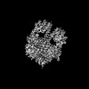
 PDBj
PDBj


 Assembly
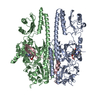

 Spodoptera frugiperda (fall armyworm) / References: UniProt: P14713

 Mass: 586.678 Da / Num. of mol.: 2 / Source method: obtained synthetically / Formula: C33H38N4O6 / Feature type: SUBJECT OF INVESTIGATION
 Sample preparation
 Electron microscopy imaging
 FIELD EMISSION GUN / Accelerating voltage: 300 kV / Illumination mode: FLOOD BEAM
 Processing