 Movie
Movie Controller


+ Open data

- Basic information
| Entry | Database: PDB / ID: 8y8v | |||||||||||||||||||||||||||||||||||||||||||||
|---|---|---|---|---|---|---|---|---|---|---|---|---|---|---|---|---|---|---|---|---|---|---|---|---|---|---|---|---|---|---|---|---|---|---|---|---|---|---|---|---|---|---|---|---|---|---|
| Title | Cryo-EM structure of AQP7 in POPC nanodisc | |||||||||||||||||||||||||||||||||||||||||||||
 Components Components | Green fluorescent protein,Aquaporin-7 | |||||||||||||||||||||||||||||||||||||||||||||
 Keywords Keywords | MEMBRANE PROTEIN / water channel / aquaporin / aquaglyceroporin / glycerol | |||||||||||||||||||||||||||||||||||||||||||||
| Function / homology |  Function and homology information Function and homology informationTransport of glycerol from adipocytes to the liver by Aquaporins / glycerol channel activity / Passive transport by Aquaporins / glycerol transmembrane transport / urea transmembrane transporter activity / water channel activity / water transport / lipid droplet / cytoplasmic vesicle membrane / bioluminescence ...Transport of glycerol from adipocytes to the liver by Aquaporins / glycerol channel activity / Passive transport by Aquaporins / glycerol transmembrane transport / urea transmembrane transporter activity / water channel activity / water transport / lipid droplet / cytoplasmic vesicle membrane / bioluminescence / generation of precursor metabolites and energy / cell-cell junction / basolateral plasma membrane / plasma membrane / cytoplasm Similarity search - Function | |||||||||||||||||||||||||||||||||||||||||||||
| Biological species |   Aequorea victoria (jellyfish) Aequorea victoria (jellyfish) Homo sapiens (human) Homo sapiens (human) | |||||||||||||||||||||||||||||||||||||||||||||
| Method | ELECTRON MICROSCOPY / single particle reconstruction / cryo EM / Resolution: 2.49 Å | |||||||||||||||||||||||||||||||||||||||||||||
 Authors Authors | Kozai, D. / Suzuki, S. / Kamegawa, A. / Nishikawa, K. / Suzuki, H. / Fujiyosh, Y. | |||||||||||||||||||||||||||||||||||||||||||||
| Funding support |  Japan, 2items Japan, 2items
| |||||||||||||||||||||||||||||||||||||||||||||
 Citation Citation | Journal: Nat Commun / Year: 2025 Title: Narrowed pore conformations of aquaglyceroporins AQP3 and GlpF. Authors: Daisuke Kozai / Masao Inoue / Shota Suzuki / Akiko Kamegawa / Kouki Nishikawa / Hiroshi Suzuki / Toru Ekimoto / Mitsunori Ikeguchi / Yoshinori Fujiyoshi / Abstract: Aquaglyceroporins such as aquaporin-3 (AQP3) and its bacterial homologue GlpF facilitate water and glycerol permeation across lipid bilayers. X-ray crystal structures of GlpF showed open pore ...Aquaglyceroporins such as aquaporin-3 (AQP3) and its bacterial homologue GlpF facilitate water and glycerol permeation across lipid bilayers. X-ray crystal structures of GlpF showed open pore conformations, and AQP3 has also been predicted to adopt this conformation. Here we present cryo-electron microscopy structures of rat AQP3 and GlpF in different narrowed pore conformations. In n-dodecyl-β-D-maltopyranoside detergent micelles, aromatic/arginine constriction filter residues of AQP3 containing Tyr212 form a 2.8-Å diameter pore, whereas in 1-palmitoyl-2-oleoyl-sn-glycero-3-phosphocholine (POPC) nanodiscs, Tyr212 inserts into the pore. Molecular dynamics simulation shows the Tyr212-in conformation is stable and largely suppresses water permeability. AQP3 reconstituted in POPC liposomes exhibits water and glycerol permeability, suggesting that the Tyr212-in conformation may be altered during permeation. AQP3 Y212F and Y212T mutant structures suggest that the aromatic residue drives the pore-inserted conformation. The aromatic residue is conserved in AQP7 and GlpF, but neither structure exhibits the AQP3-like conformation in POPC nanodiscs. Unexpectedly, the GlpF pore is covered by an intracellular loop, but the loop is flexible and not primarily related to the GlpF permeability. Our findings illuminate the unique AQP3 conformation and structural diversity of aquaglyceroporins. | |||||||||||||||||||||||||||||||||||||||||||||
| History |
|
- Structure visualization
| Structure viewer | Molecule: MolmilJmol/JSmol |
|---|
- Downloads & links
-Download
| PDBx/mmCIF format | 8y8v.cif.gz | 71.2 KB | Display | PDBx/mmCIF format |
|---|---|---|---|---|
| PDB format | pdb8y8v.ent.gz | 44.4 KB | Display | PDB format |
| PDBx/mmJSON format | 8y8v.json.gz | Tree view | PDBx/mmJSON format | |
| Others |  Other downloads Other downloads |
-Validation report
| Arichive directory | https://data.pdbj.org/pub/pdb/validation_reports/y8/8y8vftp://data.pdbj.org/pub/pdb/validation_reports/y8/8y8v | HTTPS FTP |
|---|
-Related structure data
| Related structure data |  39060MC 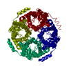 8y8nC 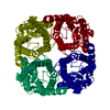 8y8oC  8y8pC 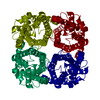 8y8qC 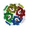 8y8rC 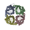 8y8sC 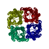 8y8wC  8y8xC M: map data used to model this data C: citing same article ( |
|---|---|
| Similar structure data |
-Links
 PDBj
PDBj


- Assembly
| Deposited unit | 
|
|---|---|
| 1 | 
|
| 2 |
|
| 3 | 
|
| Symmetry | Point symmetry: (Schoenflies symbol: C4 (4 fold cyclic)) |
-Components
| #1: Protein | Mass: 67259.117 Da / Num. of mol.: 1 Source method: isolated from a genetically manipulated source Details: 5-12 His tag 13-251 GFP tag,255-261 TEV protease digestion site Source: (gene. exp.) Aequorea victoria (jellyfish), (gene. exp.) Homo sapiens (human)Gene: GFP, AQP7, AQP7L, AQP9 / Production host:   Spodoptera frugiperda (fall armyworm) / References: UniProt: P42212, UniProt: O14520 Spodoptera frugiperda (fall armyworm) / References: UniProt: P42212, UniProt: O14520 |
|---|---|
| #2: Chemical | ChemComp-P5S /   Mass: 792.075 Da / Num. of mol.: 1 / Source method: obtained synthetically / Formula: C42H82NO10P / Feature type: SUBJECT OF INVESTIGATION Mass: 792.075 Da / Num. of mol.: 1 / Source method: obtained synthetically / Formula: C42H82NO10P / Feature type: SUBJECT OF INVESTIGATION |
| Has ligand of interest | Y |
| Has protein modification | N |
-Experimental details
-Experiment
| Experiment | Method: ELECTRON MICROSCOPY |
|---|---|
| EM experiment | Aggregation state: PARTICLE / 3D reconstruction method: single particle reconstruction |
- Sample preparation
| Component | Name: Tetramer of AQP7 in POPC nanodisc / Type: ORGANELLE OR CELLULAR COMPONENT / Entity ID: #1 / Source: RECOMBINANT | ||||||||||||
|---|---|---|---|---|---|---|---|---|---|---|---|---|---|
| Molecular weight | Experimental value: NO | ||||||||||||
| Source (natural) |
| ||||||||||||
| Source (recombinant) | Organism: Spodoptera frugiperda (fall armyworm) | ||||||||||||
| Buffer solution | pH: 8 | ||||||||||||
| Specimen | Embedding applied: NO / Shadowing applied: NO / Staining applied: NO / Vitrification applied: YES | ||||||||||||
| Vitrification | Cryogen name: ETHANE |
- Electron microscopy imaging
| Microscopy | Model: JEOL CRYO ARM 300 |
|---|---|
| Electron gun | Electron source:  FIELD EMISSION GUN / Accelerating voltage: 300 kV / Illumination mode: FLOOD BEAM FIELD EMISSION GUN / Accelerating voltage: 300 kV / Illumination mode: FLOOD BEAM |
| Electron lens | Mode: BRIGHT FIELD / Nominal defocus max: 2500 nm / Nominal defocus min: 500 nm |
| Image recording | Electron dose: 65.3 e/Å2 / Detector mode: COUNTING / Film or detector model: GATAN K2 SUMMIT (4k x 4k) |
- Processing
| EM software | Name: PHENIX / Category: model refinement | ||||||||||||||||||||||||
|---|---|---|---|---|---|---|---|---|---|---|---|---|---|---|---|---|---|---|---|---|---|---|---|---|---|
| CTF correction | Type: PHASE FLIPPING AND AMPLITUDE CORRECTION | ||||||||||||||||||||||||
| Symmetry | Point symmetry: C4 (4 fold cyclic) | ||||||||||||||||||||||||
| 3D reconstruction | Resolution: 2.49 Å / Resolution method: FSC 0.143 CUT-OFF / Num. of particles: 148023 / Symmetry type: POINT | ||||||||||||||||||||||||
| Refinement | Highest resolution: 2.49 Å Stereochemistry target values: REAL-SPACE (WEIGHTED MAP SUM AT ATOM CENTERS) | ||||||||||||||||||||||||
| Refine LS restraints |
|
