 Movie
Movie Controller

[English] 日本語
 Yorodumi
- PDB-8y1d: 2up-TM conformation of HKU1-B S protein after incubation of the r... -
+ Open data

- Basic information
| Entry | Database: PDB / ID: 8y1d | ||||||
|---|---|---|---|---|---|---|---|
| Title | 2up-TM conformation of HKU1-B S protein after incubation of the receptor | ||||||
 Components Components |
| ||||||
 Keywords Keywords | VIRAL PROTEIN/HYDROLASE / HCoV-HKU1 / VIRAL PROTEIN-HYDROLASE complex | ||||||
| Function / homology |  Function and homology information Function and homology informationtransmembrane protease serine 2 / protein autoprocessing / Attachment and Entry / serine-type peptidase activity / viral translation / Induction of Cell-Cell Fusion / entry receptor-mediated virion attachment to host cell / Attachment and Entry / host cell endoplasmic reticulum-Golgi intermediate compartment membrane / positive regulation of viral entry into host cell ...transmembrane protease serine 2 / protein autoprocessing / Attachment and Entry / serine-type peptidase activity / viral translation / Induction of Cell-Cell Fusion / entry receptor-mediated virion attachment to host cell / Attachment and Entry / host cell endoplasmic reticulum-Golgi intermediate compartment membrane / positive regulation of viral entry into host cell / receptor-mediated virion attachment to host cell / endocytosis involved in viral entry into host cell / fusion of virus membrane with host plasma membrane / serine-type endopeptidase activity / fusion of virus membrane with host endosome membrane / viral envelope / host cell plasma membrane / virion membrane / proteolysis / extracellular exosome / extracellular region / nucleoplasm / membrane / plasma membrane Similarity search - Function | ||||||
| Biological species |  Human coronavirus HKU1 Human coronavirus HKU1 Homo sapiens (human) Homo sapiens (human) | ||||||
| Method | ELECTRON MICROSCOPY / single particle reconstruction / cryo EM / Resolution: 2.7 Å | ||||||
 Authors Authors | Xia, L.Y. / Zhang, Y.Y. / Zhou, Q. | ||||||
| Funding support |  China, 1items China, 1items
| ||||||
 Citation Citation | Journal: Cell Res / Year: 2024 Title: Structural basis for the recognition of HCoV-HKU1 by human TMPRSS2. Authors: Lingyun Xia / Yuanyuan Zhang / Qiang Zhou / | ||||||
| History |
|
- Structure visualization
| Structure viewer | Molecule: MolmilJmol/JSmol |
|---|
- Downloads & links
-Download
| PDBx/mmCIF format | 8y1d.cif.gz | 776.5 KB | Display | PDBx/mmCIF format |
|---|---|---|---|---|
| PDB format | pdb8y1d.ent.gz | 631.7 KB | Display | PDB format |
| PDBx/mmJSON format | 8y1d.json.gz | Tree view | PDBx/mmJSON format | |
| Others |  Other downloads Other downloads |
-Validation report
| Arichive directory | https://data.pdbj.org/pub/pdb/validation_reports/y1/8y1dftp://data.pdbj.org/pub/pdb/validation_reports/y1/8y1d | HTTPS FTP |
|---|
-Related structure data
| Related structure data |  38832MC  8y19C 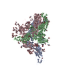 8y1aC  8y1bC  8y1cC  8y1eC 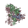 8y1fC 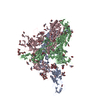 8y1gC 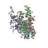 8y1hC M: map data used to model this data C: citing same article ( |
|---|---|
| Similar structure data |


-Links
 PDBj
PDBj


- Assembly
| Deposited unit | 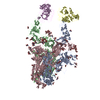
|
|---|---|
| 1 |
|
-Components
| #1: Protein | Mass: 143475.344 Da / Num. of mol.: 3 Source method: isolated from a genetically manipulated source Source: (gene. exp.) Human coronavirus HKU1 (isolate N2) / Gene: S, 3 / Production host: Homo sapiens (human) / References: UniProt: Q14EB0#2: Protein | Mass: 42368.785 Da / Num. of mol.: 2 Source method: isolated from a genetically manipulated source Source: (gene. exp.) Homo sapiens (human) / Gene: TMPRSS2, PRSS10 / Production host: Homo sapiens (human)References: UniProt: O15393, transmembrane protease serine 2 #3: Polysaccharide | Source method: isolated from a genetically manipulated source #4: Polysaccharide | 2-acetamido-2-deoxy-beta-D-glucopyranose-(1-4)-2-acetamido-2-deoxy-beta-D-glucopyranose Source method: isolated from a genetically manipulated source #5: Sugar | ChemComp-NAG /   Type: D-saccharide, beta linking / Mass: 221.208 Da / Num. of mol.: 51 Type: D-saccharide, beta linking / Mass: 221.208 Da / Num. of mol.: 51Source method: isolated from a genetically manipulated source Formula: C8H15NO6 / Feature type: SUBJECT OF INVESTIGATION Has ligand of interest | Y | Has protein modification | Y | |
|---|
-Experimental details
-Experiment
| Experiment | Method: ELECTRON MICROSCOPY |
|---|---|
| EM experiment | Aggregation state: PARTICLE / 3D reconstruction method: single particle reconstruction |
- Sample preparation
| Component | Name: 2up-TM conformation of HKU1-B S protein after incubation of the receptor Type: COMPLEX / Entity ID: #1-#2 / Source: RECOMBINANT | ||||||||||||
|---|---|---|---|---|---|---|---|---|---|---|---|---|---|
| Source (natural) |
| ||||||||||||
| Source (recombinant) | Organism: Homo sapiens (human) | ||||||||||||
| Buffer solution | pH: 8 | ||||||||||||
| Specimen | Embedding applied: NO / Shadowing applied: NO / Staining applied: NO / Vitrification applied: YES | ||||||||||||
| Vitrification | Cryogen name: ETHANE |
- Electron microscopy imaging
| Experimental equipment |  Model: Titan Krios / Image courtesy: FEI Company |
|---|---|
| Microscopy | Model: FEI TITAN KRIOS |
| Electron gun | Electron source:  FIELD EMISSION GUN / Accelerating voltage: 300 kV / Illumination mode: FLOOD BEAM FIELD EMISSION GUN / Accelerating voltage: 300 kV / Illumination mode: FLOOD BEAM |
| Electron lens | Mode: BRIGHT FIELD / Nominal defocus max: 2200 nm / Nominal defocus min: 1200 nm / Alignment procedure: COMA FREE |
| Specimen holder | Cryogen: NITROGEN / Specimen holder model: FEI TITAN KRIOS AUTOGRID HOLDER |
| Image recording | Electron dose: 50 e/Å2 / Film or detector model: GATAN K3 BIOQUANTUM (6k x 4k) |
- Processing
| EM software | Name: cryoSPARC / Version: 4 / Category: 3D reconstruction |
|---|---|
| CTF correction | Type: PHASE FLIPPING AND AMPLITUDE CORRECTION |
| 3D reconstruction | Resolution: 2.7 Å / Resolution method: FSC 0.143 CUT-OFF / Num. of particles: 408603 / Symmetry type: POINT |
