 Movie
Movie Controller

[English] 日本語
 Yorodumi
- PDB-8y0h: Structure of CXCR3 in complex with VUF11418 (Receptor-ligand focu... -
+ Open data

- Basic information
| Entry | Database: PDB / ID: 8y0h | ||||||||||||||||||||||||
|---|---|---|---|---|---|---|---|---|---|---|---|---|---|---|---|---|---|---|---|---|---|---|---|---|---|
| Title | Structure of CXCR3 in complex with VUF11418 (Receptor-ligand focused map) | ||||||||||||||||||||||||
 Components Components | C-X-C chemokine receptor type 3 | ||||||||||||||||||||||||
 Keywords Keywords | SIGNALING PROTEIN / GPCR / Arrestin | ||||||||||||||||||||||||
| Function / homology |  Function and homology information Function and homology informationregulation of leukocyte migration / chemokine binding / C-X-C chemokine binding / chemokine receptor activity / C-X-C chemokine receptor activity / T cell chemotaxis / positive regulation of chemotaxis / C-C chemokine binding / C-C chemokine receptor activity / negative regulation of execution phase of apoptosis ...regulation of leukocyte migration / chemokine binding / C-X-C chemokine binding / chemokine receptor activity / C-X-C chemokine receptor activity / T cell chemotaxis / positive regulation of chemotaxis / C-C chemokine binding / C-C chemokine receptor activity / negative regulation of execution phase of apoptosis / Chemokine receptors bind chemokines / negative regulation of endothelial cell proliferation / positive regulation of execution phase of apoptosis / regulation of cell adhesion / negative regulation of angiogenesis / positive regulation of release of sequestered calcium ion into cytosol / cell chemotaxis / calcium-mediated signaling / chemotaxis / positive regulation of angiogenesis / adenylate cyclase-activating G protein-coupled receptor signaling pathway / positive regulation of cytosolic calcium ion concentration / signaling receptor activity / angiogenesis / G alpha (i) signalling events / cell surface receptor signaling pathway / cell adhesion / immune response / G protein-coupled receptor signaling pathway / inflammatory response / external side of plasma membrane / apoptotic process / positive regulation of cell population proliferation / cell surface / positive regulation of transcription by RNA polymerase II / plasma membrane / cytoplasm Similarity search - Function | ||||||||||||||||||||||||
| Biological species |  Homo sapiens (human) Homo sapiens (human) | ||||||||||||||||||||||||
| Method | ELECTRON MICROSCOPY / single particle reconstruction / cryo EM / Resolution: 3.53 Å | ||||||||||||||||||||||||
 Authors Authors | Sano, F.K. / Saha, S. / Sharma, S. / Ganguly, M. / Shihoya, W. / Nureki, O. / Shukla, A.K. / Banerjee, R. | ||||||||||||||||||||||||
| Funding support |  India, 1items India, 1items
| ||||||||||||||||||||||||
 Citation Citation | Journal: Nat Commun / Year: 2025 Title: Structural visualization of small molecule recognition by CXCR3 uncovers dual-agonism in the CXCR3-CXCR7 system. Authors: Shirsha Saha / Fumiya K Sano / Saloni Sharma / Manisankar Ganguly / Annu Dalal / Sudha Mishra / Divyanshu Tiwari / Hiroaki Akasaka / Takaaki A Kobayashi / Nabarun Roy / Nashrah Zaidi / ...Authors: Shirsha Saha / Fumiya K Sano / Saloni Sharma / Manisankar Ganguly / Annu Dalal / Sudha Mishra / Divyanshu Tiwari / Hiroaki Akasaka / Takaaki A Kobayashi / Nabarun Roy / Nashrah Zaidi / Yuzuru Itoh / Rob Leurs / Ramanuj Banerjee / Wataru Shihoya / Osamu Nureki / Arun K Shukla /   Abstract: Chemokine receptors are critically involved in multiple physiological and pathophysiological processes related to immune response mechanisms. Most chemokine receptors are prototypical GPCRs although ...Chemokine receptors are critically involved in multiple physiological and pathophysiological processes related to immune response mechanisms. Most chemokine receptors are prototypical GPCRs although some also exhibit naturally-encoded signaling-bias toward β-arrestins (βarrs). C-X-C type chemokine receptors, namely CXCR3 and CXCR7, constitute a pair wherein the former is a prototypical GPCR while the latter exhibits selective coupling to βarrs despite sharing a common natural agonist: CXCL11. Moreover, CXCR3 and CXCR7 also recognize small molecule agonists suggesting a modular orthosteric ligand binding pocket. Here, we determine cryo-EM structures of CXCR3 in an Apo-state and in complex with small molecule agonists biased toward G-proteins or βarrs. These structural snapshots uncover an allosteric network bridging the ligand-binding pocket to intracellular side, driving the transducer-coupling bias at this receptor. Furthermore, structural topology of the orthosteric binding pocket also allows us to discover and validate that selected small molecule agonists of CXCR3 display robust agonism at CXCR7. Collectively, our study offers molecular insights into signaling-bias and dual agonism in the CXCR3-CXCR7 system with therapeutic implications. | ||||||||||||||||||||||||
| History |
|
- Structure visualization
| Structure viewer | Molecule: MolmilJmol/JSmol |
|---|
- Downloads & links
-Download
| PDBx/mmCIF format | 8y0h.cif.gz | 65 KB | Display | PDBx/mmCIF format |
|---|---|---|---|---|
| PDB format | pdb8y0h.ent.gz | Display | PDB format | |
| PDBx/mmJSON format | 8y0h.json.gz | Tree view | PDBx/mmJSON format | |
| Others |  Other downloads Other downloads |
-Validation report
| Arichive directory | https://data.pdbj.org/pub/pdb/validation_reports/y0/8y0hftp://data.pdbj.org/pub/pdb/validation_reports/y0/8y0h | HTTPS FTP |
|---|
-Related structure data
| Related structure data |  38803MC  8xxyC  8xxzC  8xyiC 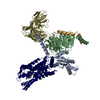 8xykC 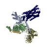 8y0nC M: map data used to model this data C: citing same article ( |
|---|---|
| Similar structure data |
-Links
 PDBj
PDBj


- Assembly
| Deposited unit | 
|
|---|---|
| 1 |
|
-Components
| #1: Protein | Mass: 46597.906 Da / Num. of mol.: 1 Source method: isolated from a genetically manipulated source Source: (gene. exp.) Homo sapiens (human) / Gene: CXCR3 / Production host:   Spodoptera frugiperda (fall armyworm) / References: UniProt: P49682 Spodoptera frugiperda (fall armyworm) / References: UniProt: P49682 |
|---|---|
| #2: Chemical | ChemComp-A1LW2 / [( Mass: 472.425 Da / Num. of mol.: 1 / Source method: obtained synthetically / Formula: C25H31IN / Feature type: SUBJECT OF INVESTIGATION |
| Has ligand of interest | Y |
| Has protein modification | Y |
-Experimental details
-Experiment
| Experiment | Method: ELECTRON MICROSCOPY |
|---|---|
| EM experiment | Aggregation state: PARTICLE / 3D reconstruction method: single particle reconstruction |
- Sample preparation
| Component | Name: C-X-C chemokine receptor type 3 in complex with VUF11418 Type: COMPLEX / Entity ID: #1 / Source: RECOMBINANT |
|---|---|
| Molecular weight | Experimental value: NO |
| Source (natural) | Organism: Homo sapiens (human) |
| Source (recombinant) | Organism: Spodoptera frugiperda (fall armyworm) |
| Buffer solution | pH: 7.4 |
| Specimen | Embedding applied: NO / Shadowing applied: NO / Staining applied: NO / Vitrification applied: YES |
| Vitrification | Cryogen name: ETHANE |
- Electron microscopy imaging
| Experimental equipment |  Model: Titan Krios / Image courtesy: FEI Company |
|---|---|
| Microscopy | Model: FEI TITAN KRIOS |
| Electron gun | Electron source:  FIELD EMISSION GUN / Accelerating voltage: 300 kV / Illumination mode: FLOOD BEAM FIELD EMISSION GUN / Accelerating voltage: 300 kV / Illumination mode: FLOOD BEAM |
| Electron lens | Mode: BRIGHT FIELD / Nominal defocus max: 1600 nm / Nominal defocus min: 800 nm |
| Specimen holder | Cryogen: NITROGEN / Specimen holder model: FEI TITAN KRIOS AUTOGRID HOLDER |
| Image recording | Electron dose: 50.1 e/Å2 / Film or detector model: GATAN K3 (6k x 4k) |
- Processing
| EM software |
| ||||||||||||||||||||
|---|---|---|---|---|---|---|---|---|---|---|---|---|---|---|---|---|---|---|---|---|---|
| CTF correction | Type: NONE | ||||||||||||||||||||
| 3D reconstruction | Resolution: 3.53 Å / Resolution method: FSC 0.143 CUT-OFF / Num. of particles: 150213 / Symmetry type: POINT | ||||||||||||||||||||
| Atomic model building | Protocol: FLEXIBLE FIT / Space: REAL | ||||||||||||||||||||
| Atomic model building | Source name: AlphaFold / Type: in silico model |
