 Movie
Movie Controller


+ Open data

- Basic information
| Entry | Database: PDB / ID: 8xzz | ||||||
|---|---|---|---|---|---|---|---|
| Title | Structure of a xylanase Xyl-1 M4 E175A in complex with xylobiose | ||||||
 Components Components | Endo-1,4-beta-xylanase | ||||||
 Keywords Keywords | HYDROLASE / Evolutionarily conserved mutations / Xylanase robustness / Non-loop region / Substrate binding patterns / Conformational dynamics | ||||||
| Function / homology |  Function and homology information Function and homology informationendo-1,4-beta-xylanase activity / endo-1,4-beta-xylanase / xylan catabolic process / extracellular region Similarity search - Function | ||||||
| Biological species |  | ||||||
| Method |  X-RAY DIFFRACTION / SYNCHROTRON / MOLECULAR REPLACEMENT / Resolution: 1.72 Å X-RAY DIFFRACTION / SYNCHROTRON / MOLECULAR REPLACEMENT / Resolution: 1.72 Å | ||||||
 Authors Authors | Xiang, W.L. / Huang, J.-W. / Yang, Y. / Chen, C.-C. / Guo, R.-T. | ||||||
| Funding support |  China, 1items China, 1items
| ||||||
 Citation Citation | Journal: J.Agric.Food Chem. / Year: 2024 Title: Unleashing the Power of Evolution in Xylanase Engineering: Investigating the Role of Distal Mutation Regulation. Authors: Wu, Y. / Yang, Y. / Lu, G. / Xiang, W.L. / Sun, T.Y. / Chen, K.W. / Lv, X. / Gui, Y.F. / Zeng, R.Q. / Du, Y.K. / Fu, C.H. / Huang, J.W. / Chen, C.C. / Guo, R.T. / Yu, L.J. | ||||||
| History |
|
- Structure visualization
| Structure viewer | Molecule: MolmilJmol/JSmol |
|---|
- Downloads & links
-Download
| PDBx/mmCIF format | 8xzz.cif.gz | 179.1 KB | Display | PDBx/mmCIF format |
|---|---|---|---|---|
| PDB format | pdb8xzz.ent.gz | 138.8 KB | Display | PDB format |
| PDBx/mmJSON format | 8xzz.json.gz | Tree view | PDBx/mmJSON format | |
| Others |  Other downloads Other downloads |
-Validation report
| Summary document | 8xzz_validation.pdf.gz | 2.5 MB | Display | wwPDB validaton report |
|---|---|---|---|---|
| Full document | 8xzz_full_validation.pdf.gz | 2.6 MB | Display | |
| Data in XML | 8xzz_validation.xml.gz | 42.7 KB | Display | |
| Data in CIF | 8xzz_validation.cif.gz | 59 KB | Display | |
| Arichive directory | https://data.pdbj.org/pub/pdb/validation_reports/xz/8xzzftp://data.pdbj.org/pub/pdb/validation_reports/xz/8xzz | HTTPS FTP |
-Related structure data
| Related structure data |  8xzxC  8xzyC  8y00C  3wp4S S: Starting model for refinement C: citing same article ( |
|---|---|
| Similar structure data |
-Links
 PDBj
PDBj
- Assembly
| Deposited unit | 
| ||||||||
|---|---|---|---|---|---|---|---|---|---|
| 1 | 
| ||||||||
| 2 | 
| ||||||||
| 3 | 
| ||||||||
| 4 | 
| ||||||||
| Unit cell |
|
-Components
| #1: Protein | Mass: 21502.773 Da / Num. of mol.: 4 / Mutation: E176A,D31P,A34Q,K118Q,A168H Source method: isolated from a genetically manipulated source Source: (gene. exp.)  #2: Polysaccharide | beta-D-xylopyranose-(1-4)-beta-D-xylopyranose | 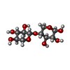  #3: Chemical | ChemComp-ZN /   Mass: 65.409 Da / Num. of mol.: 6 / Source method: obtained synthetically / Formula: Zn Mass: 65.409 Da / Num. of mol.: 6 / Source method: obtained synthetically / Formula: Zn#4: Sugar | ChemComp-XYP / 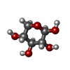  Type: D-saccharide, beta linking / Mass: 150.130 Da / Num. of mol.: 6 / Source method: isolated from a natural source / Formula: C5H10O5 / Feature type: SUBJECT OF INVESTIGATION Type: D-saccharide, beta linking / Mass: 150.130 Da / Num. of mol.: 6 / Source method: isolated from a natural source / Formula: C5H10O5 / Feature type: SUBJECT OF INVESTIGATION#5: Water | ChemComp-HOH / |  Mass: 18.015 Da / Num. of mol.: 740 / Source method: isolated from a natural source / Formula: H2O Mass: 18.015 Da / Num. of mol.: 740 / Source method: isolated from a natural source / Formula: H2OHas ligand of interest | Y | Has protein modification | N | |
|---|
-Experimental details
-Experiment
| Experiment | Method: X-RAY DIFFRACTION / Number of used crystals: 1 |
|---|
- Sample preparation
| Crystal | Density Matthews: 2.18 Å3/Da / Density % sol: 43.57 % |
|---|---|
| Crystal grow | Temperature: 295 K / Method: evaporation Details: 0.2 M sodium formate, 0.1 M Bis-Tris propane, pH 6.5, and 20% PEG 3350 |
-Data collection
| Diffraction | Mean temperature: 100 K / Serial crystal experiment: N |
|---|---|
| Diffraction source | Source: SYNCHROTRON / Site: NSRRC  / Beamline: TPS 05A / Wavelength: 0.99987 Å / Beamline: TPS 05A / Wavelength: 0.99987 Å |
| Detector | Type: DECTRIS EIGER2 X 9M / Detector: PIXEL / Date: Sep 25, 2023 |
| Radiation | Protocol: SINGLE WAVELENGTH / Monochromatic (M) / Laue (L): M / Scattering type: x-ray |
| Radiation wavelength | Wavelength: 0.99987 Å / Relative weight: 1 |
| Reflection | Resolution: 1.72→24.27 Å / Num. obs: 73843 / % possible obs: 95.5 % / Redundancy: 3.9 % / CC1/2: 0.997 / CC star: 0.999 / Rmerge(I) obs: 0.051 / Rpim(I) all: 0.031 / Rrim(I) all: 0.06 / Χ2: 1.369 / Net I/σ(I): 21.3 |
| Reflection shell | Resolution: 1.72→1.78 Å / Redundancy: 4 % / Rmerge(I) obs: 0.152 / Mean I/σ(I) obs: 13.45 / Num. unique obs: 6968 / % possible all: 90.6 |
- Processing
| Software |
| |||||||||||||||||||||||||||||||||||||||||||||||||||||||||||||||||||||||||||||||||||||||||||||||||||||||||
|---|---|---|---|---|---|---|---|---|---|---|---|---|---|---|---|---|---|---|---|---|---|---|---|---|---|---|---|---|---|---|---|---|---|---|---|---|---|---|---|---|---|---|---|---|---|---|---|---|---|---|---|---|---|---|---|---|---|---|---|---|---|---|---|---|---|---|---|---|---|---|---|---|---|---|---|---|---|---|---|---|---|---|---|---|---|---|---|---|---|---|---|---|---|---|---|---|---|---|---|---|---|---|---|---|---|---|
| Refinement | Method to determine structure: MOLECULAR REPLACEMENT Starting model: 3WP4 Resolution: 1.72→24.27 Å / SU ML: 0.17 / Cross valid method: THROUGHOUT / σ(F): 1.43 / Phase error: 19.04 / Stereochemistry target values: ML
| |||||||||||||||||||||||||||||||||||||||||||||||||||||||||||||||||||||||||||||||||||||||||||||||||||||||||
| Solvent computation | Shrinkage radii: 0.9 Å / VDW probe radii: 1.11 Å / Solvent model: FLAT BULK SOLVENT MODEL | |||||||||||||||||||||||||||||||||||||||||||||||||||||||||||||||||||||||||||||||||||||||||||||||||||||||||
| Refinement step | Cycle: LAST / Resolution: 1.72→24.27 Å
| |||||||||||||||||||||||||||||||||||||||||||||||||||||||||||||||||||||||||||||||||||||||||||||||||||||||||
| Refine LS restraints |
| |||||||||||||||||||||||||||||||||||||||||||||||||||||||||||||||||||||||||||||||||||||||||||||||||||||||||
| LS refinement shell |
|
