 Movie
Movie Controller

[English] 日本語
 Yorodumi
- PDB-8xzu: HosA transcriptional regulator from enteropathogenic Escherichia ... -
+ Open data

- Basic information
| Entry | Database: PDB / ID: 8xzu | ||||||
|---|---|---|---|---|---|---|---|
| Title | HosA transcriptional regulator from enteropathogenic Escherichia coli O127:H6 (strain E2348/69) bound with 4-hydroxy benzoic acid - Conformation II at 2.33 angstrom resolution | ||||||
 Components Components | Transcriptional regulator HosA | ||||||
 Keywords Keywords | DNA BINDING PROTEIN / Antibiotic resistance / MarR transcription factor / HosA / Enteropathogenic Escherichia coli / Paraben | ||||||
| Function / homology |  Function and homology information Function and homology information | ||||||
| Biological species |  | ||||||
| Method |  X-RAY DIFFRACTION / SYNCHROTRON / MOLECULAR REPLACEMENT / Resolution: 2.33 Å X-RAY DIFFRACTION / SYNCHROTRON / MOLECULAR REPLACEMENT / Resolution: 2.33 Å | ||||||
 Authors Authors | Manjunath, K. / Goswami, A. | ||||||
| Funding support |  India, 1items India, 1items
| ||||||
 Citation Citation | Journal: Biorxiv / Year: 2024 Title: Horizontally acquired HosA transcription factor bound with 4-hydroxy-benzoic acid exhibits unique tug-of-water dynamics. Authors: Goswami, A. / Ullah, S. / Brito, J.A. | ||||||
| History |
|
- Structure visualization
| Structure viewer | Molecule: MolmilJmol/JSmol |
|---|
- Downloads & links
-Download
| PDBx/mmCIF format | 8xzu.cif.gz | 69.8 KB | Display | PDBx/mmCIF format |
|---|---|---|---|---|
| PDB format | pdb8xzu.ent.gz | 49.6 KB | Display | PDB format |
| PDBx/mmJSON format | 8xzu.json.gz | Tree view | PDBx/mmJSON format | |
| Others |  Other downloads Other downloads |
-Validation report
| Arichive directory | https://data.pdbj.org/pub/pdb/validation_reports/xz/8xzuftp://data.pdbj.org/pub/pdb/validation_reports/xz/8xzu | HTTPS FTP |
|---|
-Related structure data


-Links
 PDBj
PDBj- Assembly
| Deposited unit | 
| ||||||||
|---|---|---|---|---|---|---|---|---|---|
| 1 | 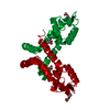
| ||||||||
| Unit cell |
|
-Components
| #1: Protein | Mass: 16592.092 Da / Num. of mol.: 1 Source method: isolated from a genetically manipulated source Details: HosA protein Source: (gene. exp.) Gene: hosA / Production host: |
|---|---|
| #2: Chemical | ChemComp-PHB / 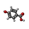  Mass: 138.121 Da / Num. of mol.: 1 / Source method: obtained synthetically / Formula: C7H6O3 Mass: 138.121 Da / Num. of mol.: 1 / Source method: obtained synthetically / Formula: C7H6O3 |
| #3: Chemical | ChemComp-PEG /   Mass: 106.120 Da / Num. of mol.: 1 / Source method: obtained synthetically / Formula: C4H10O3 Mass: 106.120 Da / Num. of mol.: 1 / Source method: obtained synthetically / Formula: C4H10O3 |
| #4: Water | ChemComp-HOH /  Mass: 18.015 Da / Num. of mol.: 28 / Source method: isolated from a natural source / Formula: H2O Mass: 18.015 Da / Num. of mol.: 28 / Source method: isolated from a natural source / Formula: H2O |
| Has ligand of interest | N |
-Experimental details
-Experiment
| Experiment | Method: X-RAY DIFFRACTION / Number of used crystals: 1 |
|---|
- Sample preparation
| Crystal | Density Matthews: 3.26 Å3/Da / Density % sol: 62.24 % / Description: Rod shaped |
|---|---|
| Crystal grow | Temperature: 277.15 K / Method: microbatch / pH: 6.2 Details: Sodium phosphate dibasic/ Potassium phosphate monobasic, Sodium chloride, PEG 200 |
-Data collection
| Diffraction | Mean temperature: 100 K / Serial crystal experiment: N |
|---|---|
| Diffraction source | Source: SYNCHROTRON / Site: RRCAT INDUS-2 / Beamline: PX-BL21 / Wavelength: 0.97893 Å |
| Detector | Type: MARMOSAIC 225 mm CCD / Detector: CCD / Date: Sep 13, 2023 |
| Radiation | Protocol: SINGLE WAVELENGTH / Monochromatic (M) / Laue (L): M / Scattering type: x-ray |
| Radiation wavelength | Wavelength: 0.97893 Å / Relative weight: 1 |
| Reflection | Resolution: 2.33→67.29 Å / Num. obs: 9878 / % possible obs: 100 % / Redundancy: 8.8 % / Biso Wilson estimate: 33.527 Å2 / CC1/2: 0.997 / Rmerge(I) obs: 0.17 / Rpim(I) all: 0.06 / Rrim(I) all: 0.18 / Χ2: 0.92 / Net I/av σ(I): 10.5 / Net I/σ(I): 10.5 |
| Reflection shell | Resolution: 2.33→2.41 Å / Redundancy: 8.5 % / Rmerge(I) obs: 1.524 / Mean I/σ(I) obs: 1.5 / Num. unique obs: 933 / CC1/2: 0.543 / Rpim(I) all: 0.545 / Rrim(I) all: 1.621 / Χ2: 0.86 |
- Processing
| Software |
| ||||||||||||||||||||||||||||||||||||||||||||||||||||||||||||||||||||||||||||||||||||||||||||||||||||
|---|---|---|---|---|---|---|---|---|---|---|---|---|---|---|---|---|---|---|---|---|---|---|---|---|---|---|---|---|---|---|---|---|---|---|---|---|---|---|---|---|---|---|---|---|---|---|---|---|---|---|---|---|---|---|---|---|---|---|---|---|---|---|---|---|---|---|---|---|---|---|---|---|---|---|---|---|---|---|---|---|---|---|---|---|---|---|---|---|---|---|---|---|---|---|---|---|---|---|---|---|---|
| Refinement | Method to determine structure: MOLECULAR REPLACEMENT / Resolution: 2.33→67.29 Å / SU ML: 0.27 / Cross valid method: FREE R-VALUE / σ(F): 1.35 / Phase error: 26.15 / Stereochemistry target values: ML Details: TLS refinement and pdb redo were used in intermediate steps of refinement. Last refinement was from Phenix.refine only.
| ||||||||||||||||||||||||||||||||||||||||||||||||||||||||||||||||||||||||||||||||||||||||||||||||||||
| Solvent computation | Shrinkage radii: 0.9 Å / VDW probe radii: 1.11 Å / Solvent model: FLAT BULK SOLVENT MODEL | ||||||||||||||||||||||||||||||||||||||||||||||||||||||||||||||||||||||||||||||||||||||||||||||||||||
| Displacement parameters | Biso mean: 40.41 Å2 | ||||||||||||||||||||||||||||||||||||||||||||||||||||||||||||||||||||||||||||||||||||||||||||||||||||
| Refinement step | Cycle: LAST / Resolution: 2.33→67.29 Å
| ||||||||||||||||||||||||||||||||||||||||||||||||||||||||||||||||||||||||||||||||||||||||||||||||||||
| Refine LS restraints |
| ||||||||||||||||||||||||||||||||||||||||||||||||||||||||||||||||||||||||||||||||||||||||||||||||||||
| LS refinement shell |
| ||||||||||||||||||||||||||||||||||||||||||||||||||||||||||||||||||||||||||||||||||||||||||||||||||||
| Refinement TLS params. | Method: refined / Refine-ID: X-RAY DIFFRACTION
| ||||||||||||||||||||||||||||||||||||||||||||||||||||||||||||||||||||||||||||||||||||||||||||||||||||
| Refinement TLS group |
|
