 Movie
Movie Controller


+ Open data

- Basic information
| Entry | Database: PDB / ID: 8xzb | ||||||
|---|---|---|---|---|---|---|---|
| Title | The structure of fox ACE2 and SARS-CoV RBD complex | ||||||
 Components Components |
| ||||||
 Keywords Keywords | VIRAL PROTEIN / fox ACE2 SARS-CoV RBD | ||||||
| Function / homology |  Function and homology information Function and homology informationMaturation of spike protein / Translation of Structural Proteins / Virion Assembly and Release / ceramide biosynthetic process / Hydrolases; Acting on peptide bonds (peptidases) / positive regulation of L-proline import across plasma membrane / angiotensin-mediated drinking behavior / positive regulation of gap junction assembly / regulation of cardiac conduction / peptidyl-dipeptidase activity ...Maturation of spike protein / Translation of Structural Proteins / Virion Assembly and Release / ceramide biosynthetic process / Hydrolases; Acting on peptide bonds (peptidases) / positive regulation of L-proline import across plasma membrane / angiotensin-mediated drinking behavior / positive regulation of gap junction assembly / regulation of cardiac conduction / peptidyl-dipeptidase activity / transporter activator activity / carboxypeptidase activity / Attachment and Entry / brush border membrane / metallopeptidase activity / SARS-CoV-1 activates/modulates innate immune responses / virus receptor activity / regulation of inflammatory response / endopeptidase activity / symbiont-mediated-mediated suppression of host tetherin activity / positive regulation of viral entry into host cell / membrane fusion / host cell endoplasmic reticulum-Golgi intermediate compartment membrane / entry receptor-mediated virion attachment to host cell / receptor-mediated virion attachment to host cell / host cell surface receptor binding / symbiont-mediated suppression of host innate immune response / cilium / endocytosis involved in viral entry into host cell / fusion of virus membrane with host plasma membrane / fusion of virus membrane with host endosome membrane / viral envelope / host cell plasma membrane / virion membrane / cell surface / negative regulation of transcription by RNA polymerase II / proteolysis / : / membrane / metal ion binding / identical protein binding / cytoplasm Similarity search - Function | ||||||
| Biological species |    Severe acute respiratory syndrome-related coronavirus Severe acute respiratory syndrome-related coronavirus | ||||||
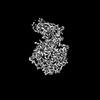
| Method | ELECTRON MICROSCOPY / single particle reconstruction / cryo EM / Resolution: 3.12 Å | ||||||
 Authors Authors | sun, J.Q. | ||||||
| Funding support |  China, 1items China, 1items
| ||||||
 Citation Citation | Journal: Virol Sin / Year: 2024 Title: The binding and structural basis of fox ACE2 to RBDs from different sarbecoviruses. Authors: Junsen Chen / Junqing Sun / Zepeng Xu / Linjie Li / Xinrui Kang / Chunliang Luo / Qi Wang / Xueyang Guo / Yan Li / Kefang Liu / Ying Wu / Abstract: Foxes are susceptible to SARS-CoV-2 in laboratory settings, and there have also been reports of natural infections of both SARS-CoV and SARS-CoV-2 in foxes. In this study, we assessed the binding ...Foxes are susceptible to SARS-CoV-2 in laboratory settings, and there have also been reports of natural infections of both SARS-CoV and SARS-CoV-2 in foxes. In this study, we assessed the binding capacities of fox ACE2 to important sarbecoviruses, including SARS-CoV, SARS-CoV-2, and animal-origin SARS-CoV-2 related viruses. Our findings demonstrated that fox ACE2 exhibits broad binding capabilities to receptor-binding domains (RBDs) of sarbecoviruses. We further determined the cryo-EM structures of fox ACE2 complexed with RBDs of SARS-CoV, SARS-CoV-2 prototype (PT), and Omicron BF.7. Through structural analysis, we identified that the K417 mutation can weaken the ability of SARS-CoV-2 sub-variants to bind to fox ACE2, thereby reducing the susceptibility of foxes to SARS-CoV-2 sub-variants. In addition, the Y498 residue in the SARS-CoV RBD plays a crucial role in forming a vital cation-π interaction with K353 in the fox ACE2 receptor. This interaction is the primary determinant for the higher affinity of the SARS-CoV RBD compared to that of the SARS-CoV-2 PT RBD. These results indicate that foxes serve as potential hosts for numerous sarbecoviruses, highlighting the critical importance of surveillance efforts. | ||||||
| History |
|
- Structure visualization
| Structure viewer | Molecule: MolmilJmol/JSmol |
|---|
- Downloads & links
-Download
| PDBx/mmCIF format | 8xzb.cif.gz | 193.7 KB | Display | PDBx/mmCIF format |
|---|---|---|---|---|
| PDB format | pdb8xzb.ent.gz | 145.7 KB | Display | PDB format |
| PDBx/mmJSON format | 8xzb.json.gz | Tree view | PDBx/mmJSON format | |
| Others |  Other downloads Other downloads |
-Validation report
| Arichive directory | https://data.pdbj.org/pub/pdb/validation_reports/xz/8xzbftp://data.pdbj.org/pub/pdb/validation_reports/xz/8xzb | HTTPS FTP |
|---|
-Related structure data
| Related structure data |  38792MC 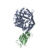 8xyzC  8xzdC M: map data used to model this data C: citing same article ( |
|---|---|
| Similar structure data |

-Links
 PDBj
PDBj

- Assembly
| Deposited unit | 
|
|---|---|
| 1 |
|
-Components
| #1: Protein | Mass: 71030.781 Da / Num. of mol.: 1 Source method: isolated from a genetically manipulated source Source: (gene. exp.)  Homo sapiens (human) Homo sapiens (human)References: UniProt: A0A3Q7RAT9, Hydrolases; Acting on peptide bonds (peptidases) |
|---|---|
| #2: Protein | Mass: 25863.234 Da / Num. of mol.: 1 / Fragment: RBD Source method: isolated from a genetically manipulated source Source: (gene. exp.) Severe acute respiratory syndrome-related coronavirusGene: S, 2 / Production host: Homo sapiens (human) / References: UniProt: P59594 |
| #3: Chemical | ChemComp-ZN /   Mass: 65.409 Da / Num. of mol.: 1 / Source method: obtained synthetically / Formula: Zn / Feature type: SUBJECT OF INVESTIGATION Mass: 65.409 Da / Num. of mol.: 1 / Source method: obtained synthetically / Formula: Zn / Feature type: SUBJECT OF INVESTIGATION |
| Has ligand of interest | Y |
| Has protein modification | Y |
-Experimental details
-Experiment
| Experiment | Method: ELECTRON MICROSCOPY |
|---|---|
| EM experiment | Aggregation state: PARTICLE / 3D reconstruction method: single particle reconstruction |
- Sample preparation
| Component | Name: The structure of fox ACE2 and PT RBD complex / Type: COMPLEX / Entity ID: #1-#2 / Source: RECOMBINANT |
|---|---|
| Source (natural) | Organism:  Severe acute respiratory syndrome coronavirus 2 Severe acute respiratory syndrome coronavirus 2 |
| Source (recombinant) | Organism: Homo sapiens (human) |
| Buffer solution | pH: 8 |
| Specimen | Conc.: 0.01 mg/ml / Embedding applied: NO / Shadowing applied: NO / Staining applied: NO / Vitrification applied: YES |
| Vitrification | Cryogen name: ETHANE |
- Electron microscopy imaging
| Experimental equipment |  Model: Titan Krios / Image courtesy: FEI Company |
|---|---|
| Microscopy | Model: FEI TITAN KRIOS |
| Electron gun | Electron source:  FIELD EMISSION GUN / Accelerating voltage: 300 kV / Illumination mode: FLOOD BEAM FIELD EMISSION GUN / Accelerating voltage: 300 kV / Illumination mode: FLOOD BEAM |
| Electron lens | Mode: BRIGHT FIELD / Nominal defocus max: 2000 nm / Nominal defocus min: 1000 nm |
| Image recording | Electron dose: 60 e/Å2 / Film or detector model: GATAN K3 (6k x 4k) |
- Processing
| CTF correction | Type: PHASE FLIPPING AND AMPLITUDE CORRECTION |
|---|---|
| 3D reconstruction | Resolution: 3.12 Å / Resolution method: FSC 0.143 CUT-OFF / Num. of particles: 345875 / Symmetry type: POINT |
