 Movie
Movie Controller

[English] 日本語
 Yorodumi
- PDB-8xq3: Structure of Nipah virus Bangladesh string G protein ectodomain t... -
+ Open data

- Basic information
| Entry | Database: PDB / ID: 8xq3 | ||||||
|---|---|---|---|---|---|---|---|
| Title | Structure of Nipah virus Bangladesh string G protein ectodomain tetramer bound to single-domain antibody n425 at 5.87 Angstroms overall resolution | ||||||
 Components Components |
| ||||||
 Keywords Keywords | VIRAL PROTEIN / Antibody | ||||||
| Biological species |  Henipavirus nipahense Henipavirus nipahense Homo sapiens (human) Homo sapiens (human) | ||||||
| Method | ELECTRON MICROSCOPY / single particle reconstruction / cryo EM / Resolution: 5.87 Å | ||||||
 Authors Authors | Sun, L. / Chen, Z. / Sun, Y. / Mao, Q. | ||||||
| Funding support |  China, 1items China, 1items
| ||||||
 Citation Citation | Journal: Nat Commun / Year: 2024 Title: Fully human single-domain antibody targeting a highly conserved cryptic epitope on the Nipah virus G protein. Authors: Yulu Wang / Yifang Sun / Zhaoling Shen / Cong Wang / Jun Qian / Qiyu Mao / Yajie Wang / Wenping Song / Yu Kong / Changyou Zhan / Zhenguo Chen / Dimiter S Dimitrov / Zhenlin Yang / Shibo ...Authors: Yulu Wang / Yifang Sun / Zhaoling Shen / Cong Wang / Jun Qian / Qiyu Mao / Yajie Wang / Wenping Song / Yu Kong / Changyou Zhan / Zhenguo Chen / Dimiter S Dimitrov / Zhenlin Yang / Shibo Jiang / Fan Wu / Lu Lu / Tianlei Ying / Lei Sun / Yanling Wu /  Abstract: Nipah virus infection, one of the top priority diseases recognized by the World Health Organization, underscores the urgent need to develop effective countermeasures against potential epidemics and ...Nipah virus infection, one of the top priority diseases recognized by the World Health Organization, underscores the urgent need to develop effective countermeasures against potential epidemics and pandemics. Here, we identify a fully human single-domain antibody that targets a highly conserved cryptic epitope situated at the dimeric interface of the Nipah virus G protein (receptor binding protein, RBP), as elucidated through structures by high-resolution cryo-electron microscopy (cryo-EM). This unique binding mode disrupts the tetramerization of the G protein, consequently obstructing the activation of the F protein and inhibiting viral membrane fusion. Furthermore, our investigations reveal that this compact antibody displays enhanced permeability across the blood-brain barrier (BBB) and demonstrates superior efficacy in eliminating pseudovirus within the brain in a murine model of Nipah virus infection, particularly compared to the well-characterized antibody m102.4 in an IgG1 format. Consequently, this single-domain antibody holds promise as a therapeutic candidate to prevent Nipah virus infections and has potential implications for vaccine development. | ||||||
| History |
|
- Structure visualization
| Structure viewer | Molecule:  MolmilJmol/JSmol MolmilJmol/JSmol |
|---|
- Downloads & links
-Download
| PDBx/mmCIF format | 8xq3.cif.gz | 409.6 KB | Display | PDBx/mmCIF format |
|---|---|---|---|---|
| PDB format | pdb8xq3.ent.gz | 340.7 KB | Display | PDB format |
| PDBx/mmJSON format | 8xq3.json.gz | Tree view | PDBx/mmJSON format | |
| Others |  Other downloads Other downloads |
-Validation report
| Arichive directory | https://data.pdbj.org/pub/pdb/validation_reports/xq/8xq3ftp://data.pdbj.org/pub/pdb/validation_reports/xq/8xq3 | HTTPS FTP |
|---|
-Related structure data
| Related structure data |  38564MC  8xpsC 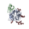 8xpyC M: map data used to model this data C: citing same article ( |
|---|

-Links
 PDBj
PDBj

- Assembly
| Deposited unit | 
|
|---|---|
| 1 |
|
-Components
-Protein / Antibody , 2 types, 8 molecules BACDFEGH
| #1: Protein | Mass: 67284.750 Da / Num. of mol.: 4 Source method: isolated from a genetically manipulated source Source: (gene. exp.) Henipavirus nipahense / Cell line (production host): Expi293F / Production host: Homo sapiens (human)#2: Antibody | Mass: 12764.128 Da / Num. of mol.: 4 Source method: isolated from a genetically manipulated source Source: (gene. exp.) Homo sapiens (human) / Cell line (production host): Expi293F / Production host: Homo sapiens (human) |
|---|
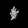
-Sugars , 4 types, 20 molecules
| #3: Polysaccharide | alpha-D-mannopyranose-(1-6)-alpha-D-mannopyranose-(1-6)-beta-D-mannopyranose-(1-4)-2-acetamido-2- ...alpha-D-mannopyranose-(1-6)-alpha-D-mannopyranose-(1-6)-beta-D-mannopyranose-(1-4)-2-acetamido-2-deoxy-beta-D-glucopyranose-(1-4)-2-acetamido-2-deoxy-beta-D-glucopyranose Source method: isolated from a genetically manipulated source #4: Polysaccharide | alpha-D-mannopyranose-(1-6)-alpha-D-mannopyranose-(1-6)-[alpha-D-mannopyranose-(1-3)]beta-D- ...alpha-D-mannopyranose-(1-6)-alpha-D-mannopyranose-(1-6)-[alpha-D-mannopyranose-(1-3)]beta-D-mannopyranose-(1-4)-2-acetamido-2-deoxy-beta-D-glucopyranose-(1-4)-2-acetamido-2-deoxy-beta-D-glucopyranose Source method: isolated from a genetically manipulated source #5: Polysaccharide | alpha-D-mannopyranose-(1-6)-beta-D-mannopyranose-(1-4)-2-acetamido-2-deoxy-beta-D-glucopyranose-(1- ...alpha-D-mannopyranose-(1-6)-beta-D-mannopyranose-(1-4)-2-acetamido-2-deoxy-beta-D-glucopyranose-(1-4)-2-acetamido-2-deoxy-beta-D-glucopyranose Source method: isolated from a genetically manipulated source #6: Polysaccharide | alpha-D-mannopyranose-(1-3)-[alpha-D-mannopyranose-(1-6)]beta-D-mannopyranose-(1-4)-2-acetamido-2- ...alpha-D-mannopyranose-(1-3)-[alpha-D-mannopyranose-(1-6)]beta-D-mannopyranose-(1-4)-2-acetamido-2-deoxy-beta-D-glucopyranose-(1-4)-2-acetamido-2-deoxy-beta-D-glucopyranose Source method: isolated from a genetically manipulated source |
|---|
-Details
| Has ligand of interest | N |
|---|---|
| Has protein modification | Y |
-Experimental details
-Experiment
| Experiment | Method: ELECTRON MICROSCOPY |
|---|---|
| EM experiment | Aggregation state: PARTICLE / 3D reconstruction method: single particle reconstruction |
- Sample preparation
| Component | Name: Nipah virus Bangladesh string G protein ectodomain tetramer in complex with single-domain antibody n425 Type: COMPLEX / Entity ID: #1-#2 / Source: RECOMBINANT |
|---|---|
| Source (natural) | Organism: Henipavirus nipahense |
| Source (recombinant) | Organism: Homo sapiens (human) |
| Buffer solution | pH: 8 |
| Specimen | Embedding applied: NO / Shadowing applied: NO / Staining applied: NO / Vitrification applied: YES |
| Vitrification | Cryogen name: ETHANE |
- Electron microscopy imaging
| Experimental equipment |  Model: Titan Krios / Image courtesy: FEI Company |
|---|---|
| Microscopy | Model: FEI TITAN KRIOS |
| Electron gun | Electron source:  FIELD EMISSION GUN / Accelerating voltage: 300 kV / Illumination mode: FLOOD BEAM FIELD EMISSION GUN / Accelerating voltage: 300 kV / Illumination mode: FLOOD BEAM |
| Electron lens | Mode: BRIGHT FIELD / Nominal defocus max: 2200 nm / Nominal defocus min: 1200 nm |
| Image recording | Electron dose: 50 e/Å2 / Film or detector model: FEI FALCON IV (4k x 4k) |
- Processing
| CTF correction | Type: PHASE FLIPPING AND AMPLITUDE CORRECTION |
|---|---|
| 3D reconstruction | Resolution: 5.87 Å / Resolution method: FSC 0.143 CUT-OFF / Num. of particles: 362285 / Symmetry type: POINT |
