[English] 日本語
Yorodumi
- PDB-8xn8: The Crystal Structure of SRC from Biortus. -

+
Open data


ID or keywords:

Loading...

-
Basic information

Entry
Database: PDB / ID: 8xn8
TitleThe Crystal Structure of SRC from Biortus.
ComponentsProto-oncogene tyrosine-protein kinase Src
KeywordsSIGNALING PROTEIN / Kinase / Transferase / Cell adhesion / ATP-binding
Function / homology
Function and homology information


Activated NTRK3 signals through PI3K / Regulation of gap junction activity / GP1b-IX-V activation signalling / Receptor Mediated Mitophagy / Nuclear signaling by ERBB4 / Signaling by EGFR / DCC mediated attractive signaling / VEGFR2 mediated cell proliferation / PECAM1 interactions / Ephrin signaling ...Activated NTRK3 signals through PI3K / Regulation of gap junction activity / GP1b-IX-V activation signalling / Receptor Mediated Mitophagy / Nuclear signaling by ERBB4 / Signaling by EGFR / DCC mediated attractive signaling / VEGFR2 mediated cell proliferation / PECAM1 interactions / Ephrin signaling / MET activates PTK2 signaling / Signaling by ERBB2 / Regulation of KIT signaling / GAB1 signalosome / Co-inhibition by CTLA4 / EPHA-mediated growth cone collapse / Co-stimulation by CD28 / Downregulation of ERBB4 signaling / Downstream signal transduction / EPH-ephrin mediated repulsion of cells / Spry regulation of FGF signaling / EPHB-mediated forward signaling / Signaling by SCF-KIT / Regulation of RUNX3 expression and activity / FCGR activation / Thrombin signalling through proteinase activated receptors (PARs) / Cyclin D associated events in G1 / RAF activation / regulation of caveolin-mediated endocytosis / regulation of toll-like receptor 3 signaling pathway / GRB2:SOS provides linkage to MAPK signaling for Integrins / regulation of cell projection assembly / positive regulation of platelet-derived growth factor receptor-beta signaling pathway / Integrin signaling / cellular response to prolactin / p130Cas linkage to MAPK signaling for integrins / positive regulation of male germ cell proliferation / MAP2K and MAPK activation / dendritic filopodium / Recycling pathway of L1 / Extra-nuclear estrogen signaling / CLEC7A (Dectin-1) signaling / negative regulation of telomere maintenance / response to mineralocorticoid / PIP3 activates AKT signaling / RHO GTPases Activate Formins / positive regulation of dephosphorylation / G alpha (i) signalling events / positive regulation of protein transport / cellular response to progesterone stimulus / BMP receptor binding / PI5P, PP2A and IER3 Regulate PI3K/AKT Signaling / negative regulation of focal adhesion assembly / positive regulation of protein processing / skeletal muscle cell proliferation / ADP signalling through P2Y purinoceptor 1 / RET signaling / regulation of intracellular estrogen receptor signaling pathway / intestinal epithelial cell development / negative regulation of neutrophil activation / positive regulation of glucose metabolic process / transcytosis / osteoclast development / connexin binding / VEGFA-VEGFR2 Pathway / focal adhesion assembly / cellular response to fluid shear stress / adherens junction organization / branching involved in mammary gland duct morphogenesis / response to acidic pH / positive regulation of Ras protein signal transduction / positive regulation of podosome assembly / positive regulation of lamellipodium morphogenesis / myoblast proliferation / cellular response to peptide hormone stimulus / negative regulation of mitochondrial depolarization / podosome / odontogenesis / cellular response to fatty acid / postsynaptic specialization, intracellular component / regulation of early endosome to late endosome transport / phospholipase activator activity / phosphorylation / oogenesis / interleukin-6-mediated signaling pathway / positive regulation of smooth muscle cell migration / DNA biosynthetic process / positive regulation of Notch signaling pathway / stress fiber assembly / negative regulation of intrinsic apoptotic signaling pathway / uterus development / regulation of cell-cell adhesion / phospholipase binding / regulation of heart rate by cardiac conduction / neurotrophin TRK receptor signaling pathway / dendritic growth cone / platelet-derived growth factor receptor signaling pathway / negative regulation of anoikis / positive regulation of epithelial cell migration / immune system process
Similarity search - Function
: / SH3 domain / SH2 domain / Src homology 2 (SH2) domain profile. / Src homology 2 domains / SH2 domain / SH2 domain superfamily / Src homology 3 domains / SH3-like domain superfamily / Src homology 3 (SH3) domain profile. ...: / SH3 domain / SH2 domain / Src homology 2 (SH2) domain profile. / Src homology 2 domains / SH2 domain / SH2 domain superfamily / Src homology 3 domains / SH3-like domain superfamily / Src homology 3 (SH3) domain profile. / SH3 domain / Tyrosine-protein kinase, catalytic domain / Tyrosine kinase, catalytic domain / Tyrosine protein kinases specific active-site signature. / Tyrosine-protein kinase, active site / Serine-threonine/tyrosine-protein kinase, catalytic domain / Protein tyrosine and serine/threonine kinase / Protein kinase, ATP binding site / Protein kinases ATP-binding region signature. / Protein kinase domain profile. / Protein kinase domain / Protein kinase-like domain superfamily
Similarity search - Domain/homology
ADENOSINE-5'-DIPHOSPHATE / Proto-oncogene tyrosine-protein kinase Src
Similarity search - Component
Biological speciesMus musculus (house mouse)
MethodX-RAY DIFFRACTION / SYNCHROTRON / MOLECULAR REPLACEMENT / Resolution: 1.95 Å
AuthorsWang, F. / Cheng, W. / Yuan, Z. / Lin, D. / Bao, C.
Funding support1items
OrganizationGrant numberCountry
Not funded
CitationJournal: To Be Published
Title: The Crystal Structure of SRC from Biortus.
Authors: Wang, F. / Cheng, W. / Yuan, Z. / Lin, D. / Bao, C.
History
DepositionDec 29, 2023Deposition site: PDBJ / Processing site: PDBJ
Revision 1.0Jan 24, 2024Provider: repository / Type: Initial release
Revision 1.1Nov 13, 2024Group: Structure summary
Category: pdbx_contact_author / pdbx_entry_details / pdbx_modification_feature
Item: _pdbx_contact_author.email / _pdbx_entry_details.has_protein_modification

-
Structure visualization

Structure viewerMolecule:
MolmilJmol/JSmol

Downloads & links

-
Assembly

Deposited unit
A: Proto-oncogene tyrosine-protein kinase Src
hetero molecules


Theoretical massNumber of molelcules
Total (without water)52,2275
Polymers51,6211
Non-polymers6064
Water7,458414
1


  • Idetical with deposited unit
  • defined by author&software
  • Evidence: gel filtration
TypeNameSymmetry operationNumber
identity operation1_555x,y,z1
Buried area940 Å2
ΔGint-16 kcal/mol
Surface area21670 Å2
MethodPISA
Unit cell
Length a, b, c (Å)51.204, 87.468, 105.802
Angle α, β, γ (deg.)90.000, 90.000, 90.000
Int Tables number19
Space group name H-MP212121

-
Components

-
Protein , 1 types, 1 molecules A

#1: Protein Proto-oncogene tyrosine-protein kinase Src / Proto-oncogene c-Src / pp60c-src / p60-Src


Mass: 51621.395 Da / Num. of mol.: 1
Source method: isolated from a genetically manipulated source
Source: (gene. exp.) Mus musculus (house mouse) / Gene: Src / Production host: 
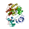
Spodoptera frugiperda (fall armyworm)
References: UniProt: P05480, non-specific protein-tyrosine kinase

-
Non-polymers , 5 types, 418 molecules

#2: Chemical ChemComp-ADP / ADENOSINE-5'-DIPHOSPHATE


Mass: 427.201 Da / Num. of mol.: 1 / Source method: obtained synthetically / Formula: C10H15N5O10P2 / Feature type: SUBJECT OF INVESTIGATION / Comment: ADP, energy-carrying molecule*YM
#3: Chemical ChemComp-MG / MAGNESIUM ION


Mass: 24.305 Da / Num. of mol.: 1 / Source method: obtained synthetically / Formula: Mg / Feature type: SUBJECT OF INVESTIGATION
#4: Chemical ChemComp-GOL / GLYCEROL / GLYCERIN / PROPANE-1,2,3-TRIOL


Mass: 92.094 Da / Num. of mol.: 1 / Source method: obtained synthetically / Formula: C3H8O3 / Feature type: SUBJECT OF INVESTIGATION
#5: Chemical ChemComp-EDO / 1,2-ETHANEDIOL / ETHYLENE GLYCOL


Mass: 62.068 Da / Num. of mol.: 1 / Source method: obtained synthetically / Formula: C2H6O2 / Feature type: SUBJECT OF INVESTIGATION
#6: Water ChemComp-HOH / water


Mass: 18.015 Da / Num. of mol.: 414 / Source method: isolated from a natural source / Formula: H2O

-
Details

Has ligand of interestY
Has protein modificationY

-
Experimental details

-
Experiment

ExperimentMethod: X-RAY DIFFRACTION / Number of used crystals: 1

-
Sample preparation

CrystalDensity Matthews: 2.29 Å3/Da / Density % sol: 46.4 %
Crystal growTemperature: 293 K / Method: vapor diffusion, sitting drop / Details: 0.2M KNO3 HCl pH6.9, 20% PEG 3350

-
Data collection

DiffractionMean temperature: 100 K / Serial crystal experiment: N
Diffraction sourceSource: SYNCHROTRON / Site: CLSI / Beamline: 08ID-1 / Wavelength: 0.953702 Å
DetectorType: DECTRIS EIGER X 9M / Detector: PIXEL / Date: Jul 29, 2023
RadiationProtocol: SINGLE WAVELENGTH / Monochromatic (M) / Laue (L): M / Scattering type: x-ray
Radiation wavelengthWavelength: 0.953702 Å / Relative weight: 1
ReflectionResolution: 1.95→46.133 Å / Num. obs: 35414 / % possible obs: 100 % / Redundancy: 9.2 % / Rmerge(I) obs: 0.133 / Net I/σ(I): 12.3
Reflection shellResolution: 1.95→2 Å / Rmerge(I) obs: 0.986 / Num. unique obs: 2453

-
Processing

Software
NameVersionClassification
REFMAC5.8.0267refinement
XDSdata reduction
Aimlessdata scaling
PHASERphasing
RefinementMethod to determine structure: MOLECULAR REPLACEMENT / Resolution: 1.95→46.133 Å / Cor.coef. Fo:Fc: 0.956 / Cor.coef. Fo:Fc free: 0.937 / WRfactor Rfree: 0.212 / WRfactor Rwork: 0.176 / SU B: 4.127 / SU ML: 0.115 / Average fsc free: 0.9207 / Average fsc work: 0.9332 / Cross valid method: FREE R-VALUE / ESU R: 0.178 / ESU R Free: 0.155
Details: Hydrogens have been added in their riding positions
RfactorNum. reflection% reflection
Rfree0.2242 1682 4.75 %
Rwork0.1847 33732 -
all0.187 --
obs-35414 99.977 %
Solvent computationIon probe radii: 0.8 Å / Shrinkage radii: 0.8 Å / VDW probe radii: 1.2 Å / Solvent model: MASK BULK SOLVENT
Displacement parametersBiso mean: 28.088 Å2
Baniso -1Baniso -2Baniso -3
1--0.28 Å2-0 Å2-0 Å2
2---0.153 Å20 Å2
3---0.432 Å2
Refinement stepCycle: LAST / Resolution: 1.95→46.133 Å
ProteinNucleic acidLigandSolventTotal
Num. atoms3620 0 38 414 4072
Refine LS restraints
Refine-IDTypeDev idealDev ideal targetNumber
X-RAY DIFFRACTIONr_bond_refined_d0.0050.0133781
X-RAY DIFFRACTIONr_bond_other_d0.0010.0173513
X-RAY DIFFRACTIONr_angle_refined_deg1.2971.6565129
X-RAY DIFFRACTIONr_angle_other_deg1.2541.5778108
X-RAY DIFFRACTIONr_dihedral_angle_1_deg6.8935461
X-RAY DIFFRACTIONr_dihedral_angle_2_deg30.27821.854205
X-RAY DIFFRACTIONr_dihedral_angle_3_deg13.47615648
X-RAY DIFFRACTIONr_dihedral_angle_4_deg15.2161528
X-RAY DIFFRACTIONr_chiral_restr0.0560.2479
X-RAY DIFFRACTIONr_gen_planes_refined0.0050.024249
X-RAY DIFFRACTIONr_gen_planes_other0.0010.02894
X-RAY DIFFRACTIONr_nbd_refined0.1920.2749
X-RAY DIFFRACTIONr_symmetry_nbd_other0.1680.23396
X-RAY DIFFRACTIONr_nbtor_refined0.1670.21819
X-RAY DIFFRACTIONr_symmetry_nbtor_other0.0730.21772
X-RAY DIFFRACTIONr_xyhbond_nbd_refined0.1730.2365
X-RAY DIFFRACTIONr_symmetry_xyhbond_nbd_other0.1930.21
X-RAY DIFFRACTIONr_symmetry_nbd_refined0.2110.217
X-RAY DIFFRACTIONr_nbd_other0.2070.254
X-RAY DIFFRACTIONr_symmetry_xyhbond_nbd_refined0.1590.225
X-RAY DIFFRACTIONr_mcbond_it1.8052.7811811
X-RAY DIFFRACTIONr_mcbond_other1.8042.7791810
X-RAY DIFFRACTIONr_mcangle_it2.9384.1622265
X-RAY DIFFRACTIONr_mcangle_other2.9374.1652266
X-RAY DIFFRACTIONr_scbond_it2.1613.0661970
X-RAY DIFFRACTIONr_scbond_other2.163.0661971
X-RAY DIFFRACTIONr_scangle_it3.5614.4852858
X-RAY DIFFRACTIONr_scangle_other3.564.4852859
X-RAY DIFFRACTIONr_lrange_it5.68433.2474457
X-RAY DIFFRACTIONr_lrange_other5.56232.7684355
LS refinement shell
Resolution (Å)Rfactor RfreeNum. reflection RfreeRfactor RworkNum. reflection RworkRefine-ID% reflection obs (%)
1.95-2.0010.3111140.2592453X-RAY DIFFRACTION100
2.001-2.0550.2541190.2342414X-RAY DIFFRACTION100
2.055-2.1150.2361030.2152339X-RAY DIFFRACTION100
2.115-2.180.241220.2062255X-RAY DIFFRACTION99.9159
2.18-2.2510.2611170.2082194X-RAY DIFFRACTION100
2.251-2.330.2531160.1972120X-RAY DIFFRACTION100
2.33-2.4180.2391070.1892046X-RAY DIFFRACTION100
2.418-2.5170.252810.1831992X-RAY DIFFRACTION100
2.517-2.6280.24850.1781911X-RAY DIFFRACTION100
2.628-2.7570.2561040.1951816X-RAY DIFFRACTION100
2.757-2.9050.277890.1921736X-RAY DIFFRACTION99.9452
2.905-3.0810.211900.1821634X-RAY DIFFRACTION100
3.081-3.2940.239860.1791552X-RAY DIFFRACTION100
3.294-3.5570.221680.1761454X-RAY DIFFRACTION100
3.557-3.8950.191510.1611374X-RAY DIFFRACTION100
3.895-4.3530.185660.1541223X-RAY DIFFRACTION99.9225
4.353-5.0230.182550.1581091X-RAY DIFFRACTION100
5.023-6.1430.179480.183932X-RAY DIFFRACTION100
6.143-8.6520.206450.175744X-RAY DIFFRACTION100
8.652-46.1330.158160.193453X-RAY DIFFRACTION99.1543

+
About Yorodumi

-
News

-
Feb 9, 2022. New format data for meta-information of EMDB entries

New format data for meta-information of EMDB entries

  • Version 3 of the EMDB header file is now the official format.
  • The previous official version 1.9 will be removed from the archive.

Related info.:EMDB header

External links:wwPDB to switch to version 3 of the EMDB data model

-
Aug 12, 2020. Covid-19 info

Covid-19 info

URL: https://pdbj.org/emnavi/covid19.php

New page: Covid-19 featured information page in EM Navigator.

Related info.:Covid-19 info / Mar 5, 2020. Novel coronavirus structure data

+
Mar 5, 2020. Novel coronavirus structure data

Novel coronavirus structure data

Related info.:Yorodumi Speices / Aug 12, 2020. Covid-19 info

External links:COVID-19 featured content - PDBj / Molecule of the Month (242):Coronavirus Proteases

+
Jan 31, 2019. EMDB accession codes are about to change! (news from PDBe EMDB page)

EMDB accession codes are about to change! (news from PDBe EMDB page)

  • The allocation of 4 digits for EMDB accession codes will soon come to an end. Whilst these codes will remain in use, new EMDB accession codes will include an additional digit and will expand incrementally as the available range of codes is exhausted. The current 4-digit format prefixed with “EMD-” (i.e. EMD-XXXX) will advance to a 5-digit format (i.e. EMD-XXXXX), and so on. It is currently estimated that the 4-digit codes will be depleted around Spring 2019, at which point the 5-digit format will come into force.
  • The EM Navigator/Yorodumi systems omit the EMD- prefix.

Related info.:Q: What is EMD? / ID/Accession-code notation in Yorodumi/EM Navigator

External links:EMDB Accession Codes are Changing Soon! / Contact to PDBj

+
Jul 12, 2017. Major update of PDB

Major update of PDB

  • wwPDB released updated PDB data conforming to the new PDBx/mmCIF dictionary.
  • This is a major update changing the version number from 4 to 5, and with Remediation, in which all the entries are updated.
  • In this update, many items about electron microscopy experimental information are reorganized (e.g. em_software).
  • Now, EM Navigator and Yorodumi are based on the updated data.

External links:wwPDB Remediation / Enriched Model Files Conforming to OneDep Data Standards Now Available in the PDB FTP Archive

-
Yorodumi

Thousand views of thousand structures

  • Yorodumi is a browser for structure data from EMDB, PDB, SASBDB, etc.
  • This page is also the successor to EM Navigator detail page, and also detail information page/front-end page for Omokage search.
  • The word "yorodu" (or yorozu) is an old Japanese word meaning "ten thousand". "mi" (miru) is to see.

Related info.:EMDB / PDB / SASBDB / Comparison of 3 databanks / Yorodumi Search / Aug 31, 2016. New EM Navigator & Yorodumi / Yorodumi Papers / Jmol/JSmol / Function and homology information / Changes in new EM Navigator and Yorodumi

Read more