 Movie
Movie Controller


+ Open data

- Basic information
| Entry | Database: PDB / ID: 8xkc | ||||||
|---|---|---|---|---|---|---|---|
| Title | The structure of HLA-A/Pep16 | ||||||
 Components Components |
| ||||||
 Keywords Keywords | VIRAL PROTEIN/IMMUNE SYSTEM / Complex / Peptide presentation / VIRAL PROTEIN-IMMUNE SYSTEM complex | ||||||
| Function / homology |  Function and homology information Function and homology informationantigen processing and presentation of peptide antigen via MHC class I / negative regulation of receptor binding / early endosome lumen / Nef mediated downregulation of MHC class I complex cell surface expression / DAP12 interactions / transferrin transport / cellular response to iron ion / Endosomal/Vacuolar pathway / Antigen Presentation: Folding, assembly and peptide loading of class I MHC / peptide antigen assembly with MHC class II protein complex ...antigen processing and presentation of peptide antigen via MHC class I / negative regulation of receptor binding / early endosome lumen / Nef mediated downregulation of MHC class I complex cell surface expression / DAP12 interactions / transferrin transport / cellular response to iron ion / Endosomal/Vacuolar pathway / Antigen Presentation: Folding, assembly and peptide loading of class I MHC / peptide antigen assembly with MHC class II protein complex / lumenal side of endoplasmic reticulum membrane / cellular response to iron(III) ion / negative regulation of forebrain neuron differentiation / MHC class II protein complex / antigen processing and presentation of exogenous protein antigen via MHC class Ib, TAP-dependent / ER to Golgi transport vesicle membrane / peptide antigen assembly with MHC class I protein complex / regulation of iron ion transport / regulation of erythrocyte differentiation / HFE-transferrin receptor complex / response to molecule of bacterial origin / MHC class I peptide loading complex / T cell mediated cytotoxicity / positive regulation of T cell cytokine production / antigen processing and presentation of endogenous peptide antigen via MHC class I / antigen processing and presentation of exogenous peptide antigen via MHC class II / positive regulation of immune response / MHC class I protein complex / peptide antigen binding / positive regulation of T cell activation / negative regulation of neurogenesis / positive regulation of receptor-mediated endocytosis / cellular response to nicotine / positive regulation of T cell mediated cytotoxicity / multicellular organismal-level iron ion homeostasis / specific granule lumen / phagocytic vesicle membrane / recycling endosome membrane / Interferon gamma signaling / Immunoregulatory interactions between a Lymphoid and a non-Lymphoid cell / negative regulation of epithelial cell proliferation / MHC class II protein complex binding / Modulation by Mtb of host immune system / late endosome membrane / sensory perception of smell / positive regulation of cellular senescence / tertiary granule lumen / DAP12 signaling / T cell differentiation in thymus / negative regulation of neuron projection development / ER-Phagosome pathway / protein refolding / early endosome membrane / symbiont-mediated disruption of host tissue / protein homotetramerization / Maturation of spike protein / Translation of Structural Proteins / Virion Assembly and Release / host cell surface / host extracellular space / viral translation / symbiont-mediated-mediated suppression of host tetherin activity / amyloid fibril formation / Induction of Cell-Cell Fusion / structural constituent of virion / intracellular iron ion homeostasis / membrane fusion / entry receptor-mediated virion attachment to host cell / Attachment and Entry / host cell endoplasmic reticulum-Golgi intermediate compartment membrane / learning or memory / positive regulation of viral entry into host cell / receptor-mediated virion attachment to host cell / host cell surface receptor binding / immune response / symbiont-mediated suppression of host innate immune response / receptor ligand activity / endocytosis involved in viral entry into host cell / endoplasmic reticulum lumen / Amyloid fiber formation / Golgi membrane / fusion of virus membrane with host plasma membrane / external side of plasma membrane / lysosomal membrane / focal adhesion / fusion of virus membrane with host endosome membrane / viral envelope / Neutrophil degranulation / symbiont entry into host cell / virion attachment to host cell / SARS-CoV-2 activates/modulates innate and adaptive immune responses / host cell plasma membrane / virion membrane / structural molecule activity / cell surface / endoplasmic reticulum / Golgi apparatus / protein homodimerization activity / extracellular space / extracellular exosome Similarity search - Function | ||||||
| Biological species |  Homo sapiens (human) Homo sapiens (human)  Severe acute respiratory syndrome coronavirus 2 Severe acute respiratory syndrome coronavirus 2 | ||||||
| Method |  X-RAY DIFFRACTION / SYNCHROTRON / MOLECULAR REPLACEMENT / Resolution: 2.18 Å X-RAY DIFFRACTION / SYNCHROTRON / MOLECULAR REPLACEMENT / Resolution: 2.18 Å | ||||||
 Authors Authors | Zhang, J.N. / Yue, C. / Liu, J. | ||||||
| Funding support |  China, 1items China, 1items
| ||||||
 Citation Citation | Journal: Immunohorizons / Year: 2024 Title: Uncommon P1 Anchor-featured Viral T Cell Epitope Preference within HLA-A*2601 and HLA-A*0101 Individuals. Authors: Zhang, J. / Yue, C. / Lin, Y. / Tian, J. / Guo, Y. / Zhang, D. / Guo, Y. / Ye, B. / Chai, Y. / Qi, J. / Zhao, Y. / Gao, G.F. / Sun, Z. / Liu, J. | ||||||
| History |
|
- Structure visualization
| Structure viewer | Molecule: MolmilJmol/JSmol |
|---|
- Downloads & links
-Download
| PDBx/mmCIF format | 8xkc.cif.gz | 112 KB | Display | PDBx/mmCIF format |
|---|---|---|---|---|
| PDB format | pdb8xkc.ent.gz | 69.1 KB | Display | PDB format |
| PDBx/mmJSON format | 8xkc.json.gz | Tree view | PDBx/mmJSON format | |
| Others |  Other downloads Other downloads |
-Validation report
| Arichive directory | https://data.pdbj.org/pub/pdb/validation_reports/xk/8xkcftp://data.pdbj.org/pub/pdb/validation_reports/xk/8xkc | HTTPS FTP |
|---|
-Related structure data
| Related structure data | 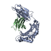 8xesC 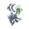 8xfzC 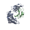 8xg2C 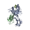 8xkeC  4nqvS S: Starting model for refinement C: citing same article ( |
|---|---|
| Similar structure data |
-Links
 PDBj
PDBj


- Assembly
| Deposited unit | 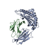
| ||||||||||||
|---|---|---|---|---|---|---|---|---|---|---|---|---|---|
| 1 |
| ||||||||||||
| Unit cell |
|
-Components
| #1: Protein | Mass: 31859.975 Da / Num. of mol.: 1 Source method: isolated from a genetically manipulated source Source: (gene. exp.) Homo sapiens (human) / Gene: HLA-A, HLA, HLA-A26 / Production host:  |
|---|---|
| #2: Protein | Mass: 11748.160 Da / Num. of mol.: 1 Source method: isolated from a genetically manipulated source Source: (gene. exp.) Homo sapiens (human) / Gene: B2M, CDABP0092, HDCMA22P / Production host: |
| #3: Protein/peptide | Mass: 1032.168 Da / Num. of mol.: 1 / Fragment: Pep16 / Source method: obtained synthetically Source: (synth.) Severe acute respiratory syndrome coronavirus 2References: UniProt: P0DTC2 |
| #4: Water | ChemComp-HOH /  Mass: 18.015 Da / Num. of mol.: 56 / Source method: isolated from a natural source / Formula: H2O Mass: 18.015 Da / Num. of mol.: 56 / Source method: isolated from a natural source / Formula: H2O |
| Has protein modification | Y |
-Experimental details
-Experiment
| Experiment | Method: X-RAY DIFFRACTION / Number of used crystals: 1 |
|---|
- Sample preparation
| Crystal | Density Matthews: 2.63 Å3/Da / Density % sol: 53.25 % |
|---|---|
| Crystal grow | Temperature: 277 K / Method: vapor diffusion, sitting drop / pH: 7 / Details: 0.1M Imidazole, 20%w/v PEG 6000 |
-Data collection
| Diffraction | Mean temperature: 100 K / Serial crystal experiment: N |
|---|---|
| Diffraction source | Source: SYNCHROTRON / Site: SSRF / Beamline: BL02U1 / Wavelength: 0.979183 Å |
| Detector | Type: DECTRIS PILATUS 6M / Detector: PIXEL / Date: Sep 8, 2023 |
| Radiation | Protocol: SINGLE WAVELENGTH / Monochromatic (M) / Laue (L): M / Scattering type: x-ray |
| Radiation wavelength | Wavelength: 0.979183 Å / Relative weight: 1 |
| Reflection | Resolution: 2.06→57.49 Å / Num. obs: 25558 / % possible obs: 99.89 % / Redundancy: 10.8 % / Biso Wilson estimate: 52.28 Å2 / Rmerge(I) obs: 0.176 / Net I/σ(I): 6.9 |
| Reflection shell | Resolution: 2.06→6.88 Å / Num. unique obs: 24245 / Rpim(I) all: 0.056 |
- Processing
| Software |
| ||||||||||||||||||||||||||||||||||||||||||||||||||||||||||||||||||||||
|---|---|---|---|---|---|---|---|---|---|---|---|---|---|---|---|---|---|---|---|---|---|---|---|---|---|---|---|---|---|---|---|---|---|---|---|---|---|---|---|---|---|---|---|---|---|---|---|---|---|---|---|---|---|---|---|---|---|---|---|---|---|---|---|---|---|---|---|---|---|---|---|
| Refinement | Method to determine structure: MOLECULAR REPLACEMENT Starting model: 4NQV Resolution: 2.18→36.28 Å / SU ML: 0.3541 / Cross valid method: FREE R-VALUE / σ(F): 1.34 / Phase error: 30.5741 Stereochemistry target values: GeoStd + Monomer Library + CDL v1.2
| ||||||||||||||||||||||||||||||||||||||||||||||||||||||||||||||||||||||
| Solvent computation | Shrinkage radii: 0.9 Å / VDW probe radii: 1.1 Å / Solvent model: FLAT BULK SOLVENT MODEL | ||||||||||||||||||||||||||||||||||||||||||||||||||||||||||||||||||||||
| Displacement parameters | Biso mean: 61.75 Å2 | ||||||||||||||||||||||||||||||||||||||||||||||||||||||||||||||||||||||
| Refinement step | Cycle: LAST / Resolution: 2.18→36.28 Å
| ||||||||||||||||||||||||||||||||||||||||||||||||||||||||||||||||||||||
| Refine LS restraints |
| ||||||||||||||||||||||||||||||||||||||||||||||||||||||||||||||||||||||
| LS refinement shell |
|
