 Movie
Movie Controller

[English] 日本語
 Yorodumi
- PDB-8xjt: Cryo-EM structure of colibactin assembly line polyketide synthase... -
+ Open data

- Basic information
| Entry | Database: PDB / ID: 8xjt | |||||||||
|---|---|---|---|---|---|---|---|---|---|---|
| Title | Cryo-EM structure of colibactin assembly line polyketide synthase ClbC (ACP-bound state) | |||||||||
 Components Components | Colibactin polyketide synthase ClbC | |||||||||
 Keywords Keywords | TRANSFERASE / colibactin / microbiome / polyketide synthase / colorectal cancer / NRPS-PKS hybrid / biosynthetic protein | |||||||||
| Function / homology |  Function and homology information Function and homology informationDIM/DIP cell wall layer assembly / fatty acid synthase activity / phosphopantetheine binding / fatty acid biosynthetic process / plasma membrane / cytoplasm Similarity search - Function | |||||||||
| Biological species |  | |||||||||
| Method | ELECTRON MICROSCOPY / single particle reconstruction / cryo EM / Resolution: 3.9 Å | |||||||||
 Authors Authors | Kim, J. / Kim, M. / Kang, J.Y. | |||||||||
| Funding support |  Korea, Republic Of, 2items Korea, Republic Of, 2items
| |||||||||
 Citation Citation | Journal: Structure / Year: 2025 Title: Structural study on human microbiome-derived polyketide synthases that assemble genotoxic colibactin. Authors: Minjae Kim / Jinwoo Kim / Gyu Sung Lee / Paul Dominic B Olinares / Yougant Airan / Jasmine L Chow / Jongseok Park / Yujin Jeong / Jiho Park / Brian T Chait / Seth B Herzon / Chung Sub Kim / Jin Young Kang /  Abstract: Colibactin, a human microbiome-derived genotoxin, promotes colorectal cancer by damaging the host gut epithelial genomes. While colibactin is synthesized via a hybrid non-ribosomal peptide synthetase ...Colibactin, a human microbiome-derived genotoxin, promotes colorectal cancer by damaging the host gut epithelial genomes. While colibactin is synthesized via a hybrid non-ribosomal peptide synthetase (NRPS)-polyketide synthase (PKS) pathway, known as pks or clb, the structural details of its biosynthetic enzymes remain limited, hindering our understanding of its biosynthesis and clinical application. In this study, we report the cryo-EM structures of two colibactin-producing PKS enzymes, ClbC and ClbI, captured in different reaction states using a substrate-mimic crosslinker. Our structural analysis revealed the binding sites of carrier protein (CP) domains of the ClbC and ClbI on their ketosynthase (KS) domains. Further, we identified a novel NRPS-PKS docking interaction between ClbI and its upstream enzyme, ClbH, mediated by the C-terminal peptide ClbH and the dimeric interface of ClbI, establishing a 1:2 stoichiometry. These findings advance our understanding of colibactin assembly line and provide broader insights into NRPS-PKS natural product biosynthesis mechanisms. | |||||||||
| History |
|
- Structure visualization
| Structure viewer | Molecule: MolmilJmol/JSmol |
|---|
- Downloads & links
-Download
| PDBx/mmCIF format | 8xjt.cif.gz | 292.5 KB | Display | PDBx/mmCIF format |
|---|---|---|---|---|
| PDB format | pdb8xjt.ent.gz | 221.2 KB | Display | PDB format |
| PDBx/mmJSON format | 8xjt.json.gz | Tree view | PDBx/mmJSON format | |
| Others |  Other downloads Other downloads |
-Validation report
| Arichive directory | https://data.pdbj.org/pub/pdb/validation_reports/xj/8xjtftp://data.pdbj.org/pub/pdb/validation_reports/xj/8xjt | HTTPS FTP |
|---|
-Related structure data
| Related structure data |  38405MC  8xblC  8xjuC  8xjyC 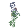 8xjzC C: citing same article ( M: map data used to model this data |
|---|---|
| Similar structure data |


-Links
 PDBj
PDBj

- Assembly
| Deposited unit | 
|
|---|---|
| 1 |
|
-Components
| #1: Protein | Mass: 99302.961 Da / Num. of mol.: 3 Source method: isolated from a genetically manipulated source Source: (gene. exp.) Gene: clbC, C1Q91_004440, EPS76_08170, EWK56_24790, FPI65_12395, H0O51_09120, HMW38_22955, IFB95_004349 Production host: Has protein modification | N | |
|---|
-Experimental details
-Experiment
| Experiment | Method: ELECTRON MICROSCOPY |
|---|---|
| EM experiment | Aggregation state: PARTICLE / 3D reconstruction method: single particle reconstruction |
- Sample preparation
| Component | Name: ACP-bound ClbC / Type: COMPLEX / Entity ID: all / Source: RECOMBINANT | |||||||||||||||||||||||||
|---|---|---|---|---|---|---|---|---|---|---|---|---|---|---|---|---|---|---|---|---|---|---|---|---|---|---|
| Source (natural) | Organism: | |||||||||||||||||||||||||
| Source (recombinant) | Organism: | |||||||||||||||||||||||||
| Buffer solution | pH: 8 | |||||||||||||||||||||||||
| Buffer component |
| |||||||||||||||||||||||||
| Specimen | Embedding applied: NO / Shadowing applied: NO / Staining applied: NO / Vitrification applied: YES | |||||||||||||||||||||||||
| Specimen support | Grid material: GOLD / Grid mesh size: 300 divisions/in. / Grid type: UltrAuFoil R1.2/1.3 | |||||||||||||||||||||||||
| Vitrification | Instrument: FEI VITROBOT MARK IV / Cryogen name: ETHANE / Humidity: 100 % / Chamber temperature: 295 K |
- Electron microscopy imaging
| Experimental equipment |  Model: Titan Krios / Image courtesy: FEI Company |
|---|---|
| Microscopy | Model: TFS KRIOS |
| Electron gun | Electron source:  FIELD EMISSION GUN / Accelerating voltage: 300 kV / Illumination mode: FLOOD BEAM FIELD EMISSION GUN / Accelerating voltage: 300 kV / Illumination mode: FLOOD BEAM |
| Electron lens | Mode: BRIGHT FIELD / Nominal magnification: 81000 X / Nominal defocus max: 2400 nm / Nominal defocus min: 800 nm / Cs: 2.73 mm |
| Specimen holder | Cryogen: NITROGEN / Specimen holder model: FEI TITAN KRIOS AUTOGRID HOLDER |
| Image recording | Electron dose: 58.85 e/Å2 / Film or detector model: GATAN K3 BIOQUANTUM (6k x 4k) / Num. of grids imaged: 2 / Num. of real images: 13070 |
| EM imaging optics | Energyfilter name: GIF Bioquantum / Energyfilter slit width: 20 eV |
| Image scans | Width: 6000 / Height: 4000 |
- Processing
| EM software |
| ||||||||||||||||||||||||||||||||||||||||
|---|---|---|---|---|---|---|---|---|---|---|---|---|---|---|---|---|---|---|---|---|---|---|---|---|---|---|---|---|---|---|---|---|---|---|---|---|---|---|---|---|---|
| CTF correction | Type: PHASE FLIPPING AND AMPLITUDE CORRECTION | ||||||||||||||||||||||||||||||||||||||||
| Particle selection | Num. of particles selected: 6827074 | ||||||||||||||||||||||||||||||||||||||||
| Symmetry | Point symmetry: C1 (asymmetric) | ||||||||||||||||||||||||||||||||||||||||
| 3D reconstruction | Resolution: 3.9 Å / Resolution method: FSC 0.143 CUT-OFF / Num. of particles: 301978 / Algorithm: FOURIER SPACE / Symmetry type: POINT | ||||||||||||||||||||||||||||||||||||||||
| Atomic model building | Protocol: RIGID BODY FIT / Space: REAL | ||||||||||||||||||||||||||||||||||||||||
| Atomic model building | Details: apo-ClbC / Source name: Other / Type: experimental model | ||||||||||||||||||||||||||||||||||||||||
| Refine LS restraints |
|
