 Movie
Movie Controller


+ Open data

- Basic information
| Entry | Database: PDB / ID: 8xip | ||||||
|---|---|---|---|---|---|---|---|
| Title | Structure of Pasireotide-SSTR1 G protein complex | ||||||
 Components Components |
| ||||||
 Keywords Keywords | MEMBRANE PROTEIN/IMMUNE SYSTEM / GPCR / SSTR1 / pasireotide / MEMBRANE PROTEIN-IMMUNE SYSTEM complex | ||||||
| Function / homology |  Function and homology information Function and homology informationsomatostatin receptor activity / neuropeptide binding / glutamate receptor signaling pathway / response to starvation / forebrain development / G protein-coupled receptor signaling pathway, coupled to cyclic nucleotide second messenger / neuropeptide signaling pathway / cerebellum development / Peptide ligand-binding receptors / cellular response to leukemia inhibitory factor ...somatostatin receptor activity / neuropeptide binding / glutamate receptor signaling pathway / response to starvation / forebrain development / G protein-coupled receptor signaling pathway, coupled to cyclic nucleotide second messenger / neuropeptide signaling pathway / cerebellum development / Peptide ligand-binding receptors / cellular response to leukemia inhibitory factor / cellular response to estradiol stimulus / Olfactory Signaling Pathway / Activation of the phototransduction cascade / G beta:gamma signalling through PLC beta / Presynaptic function of Kainate receptors / Thromboxane signalling through TP receptor / G protein-coupled acetylcholine receptor signaling pathway / Activation of G protein gated Potassium channels / Inhibition of voltage gated Ca2+ channels via Gbeta/gamma subunits / G-protein activation / G beta:gamma signalling through CDC42 / Prostacyclin signalling through prostacyclin receptor / Glucagon signaling in metabolic regulation / G beta:gamma signalling through BTK / Synthesis, secretion, and inactivation of Glucagon-like Peptide-1 (GLP-1) / ADP signalling through P2Y purinoceptor 12 / photoreceptor disc membrane / Glucagon-type ligand receptors / Sensory perception of sweet, bitter, and umami (glutamate) taste / Adrenaline,noradrenaline inhibits insulin secretion / Vasopressin regulates renal water homeostasis via Aquaporins / Glucagon-like Peptide-1 (GLP1) regulates insulin secretion / G alpha (z) signalling events / ADP signalling through P2Y purinoceptor 1 / ADORA2B mediated anti-inflammatory cytokines production / cellular response to catecholamine stimulus / G beta:gamma signalling through PI3Kgamma / adenylate cyclase-activating dopamine receptor signaling pathway / Cooperation of PDCL (PhLP1) and TRiC/CCT in G-protein beta folding / GPER1 signaling / G-protein beta-subunit binding / cellular response to prostaglandin E stimulus / heterotrimeric G-protein complex / G alpha (12/13) signalling events / Inactivation, recovery and regulation of the phototransduction cascade / extracellular vesicle / sensory perception of taste / Thrombin signalling through proteinase activated receptors (PARs) / signaling receptor complex adaptor activity / retina development in camera-type eye / GTPase binding / Ca2+ pathway / fibroblast proliferation / High laminar flow shear stress activates signaling by PIEZO1 and PECAM1:CDH5:KDR in endothelial cells / spermatogenesis / G alpha (i) signalling events / G alpha (s) signalling events / phospholipase C-activating G protein-coupled receptor signaling pathway / G alpha (q) signalling events / Ras protein signal transduction / Extra-nuclear estrogen signaling / cell population proliferation / neuron projection / G protein-coupled receptor signaling pathway / negative regulation of cell population proliferation / lysosomal membrane / GTPase activity / synapse / protein-containing complex binding / signal transduction / extracellular exosome / membrane / plasma membrane / cytoplasm / cytosol Similarity search - Function | ||||||
| Biological species |  Homo sapiens (human) Homo sapiens (human) synthetic construct (others) | ||||||
| Method | ELECTRON MICROSCOPY / single particle reconstruction / cryo EM / Resolution: 3.29 Å | ||||||
 Authors Authors | Wang, Y. / Xu, Y. / Xu, H.E. / Zhuang, Y. | ||||||
| Funding support |  China, 1items China, 1items
| ||||||
 Citation Citation | Journal: Proc Natl Acad Sci U S A / Year: 2024 Title: Selective ligand recognition and activation of somatostatin receptors SSTR1 and SSTR3. Authors: Yujue Wang / Youwei Xu / Yue Wang / Jie Zhang / Lan Chen / Xinheng He / Wenjia Fan / Kai Wu / Wen Hu / Xi Cheng / Guizhu Yang / H Eric Xu / Youwen Zhuang / Shuyang Sun / Abstract: Somatostatin receptors (SSTRs) exert critical biological functions such as negatively regulating hormone release and cell proliferation, making them popular targets for developing therapeutics to ...Somatostatin receptors (SSTRs) exert critical biological functions such as negatively regulating hormone release and cell proliferation, making them popular targets for developing therapeutics to treat endocrine disorders, especially neuroendocrine tumors. Although several panagonists mimicking the endogenous ligand somatostatin are available, the development of more effective and safer somatostatinergic therapies is limited due to a lack of molecular understanding of the ligand recognition and regulation of divergent SSTR subtypes. Here, we report four cryoelectron microscopy structures of G-coupled SSTR1 and SSTR3 activated by distinct agonists, including the FDA-approved panagonist pasireotide as well as their selective small molecule agonists L-797591 and L-796778. Our structures reveal a conserved recognition pattern of pasireotide in SSTRs attributed to the binding with a conserved extended binding pocket, distinct from SST14, octreotide, and lanreotide. Together with mutagenesis analyses, our structures further reveal the dynamic feature of ligand binding pockets in SSTR1 and SSTR3 to accommodate divergent agonists, the key determinants of ligand selectivity lying across the orthosteric pocket of different SSTR subtypes, as well as the molecular mechanism underlying diversity and conservation of receptor activation. Our work provides a framework for rational design of subtype-selective SSTR ligands and may facilitate drug development efforts targeting SSTRs with improved therapeutic efficacy and reduced side effects. | ||||||
| History |
|
- Structure visualization
| Structure viewer | Molecule: MolmilJmol/JSmol |
|---|
- Downloads & links
-Download
| PDBx/mmCIF format | 8xip.cif.gz | 212.8 KB | Display | PDBx/mmCIF format |
|---|---|---|---|---|
| PDB format | pdb8xip.ent.gz | Display | PDB format | |
| PDBx/mmJSON format | 8xip.json.gz | Tree view | PDBx/mmJSON format | |
| Others |  Other downloads Other downloads |
-Validation report
| Arichive directory | https://data.pdbj.org/pub/pdb/validation_reports/xi/8xipftp://data.pdbj.org/pub/pdb/validation_reports/xi/8xip | HTTPS FTP |
|---|
-Related structure data
| Related structure data |  38386MC 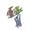 8xioC  8xiqC  8xirC M: map data used to model this data C: citing same article ( |
|---|---|
| Similar structure data |


-Links
 PDBj
PDBj


- Assembly
| Deposited unit | 
|
|---|---|
| 1 |
|
-Components
-Protein , 2 types, 2 molecules AB
| #1: Protein | Mass: 42782.043 Da / Num. of mol.: 1 / Mutation: V276Y Source method: isolated from a genetically manipulated source Source: (gene. exp.) Homo sapiens (human) / Gene: SSTR1 / Production host:  Trichoplusia ni (cabbage looper) / References: UniProt: P30872 Trichoplusia ni (cabbage looper) / References: UniProt: P30872 |
|---|---|
| #2: Protein | Mass: 41641.207 Da / Num. of mol.: 1 Source method: isolated from a genetically manipulated source Source: (gene. exp.) Homo sapiens (human) / Production host: Trichoplusia ni (cabbage looper) |
-Guanine nucleotide-binding protein ... , 2 types, 2 molecules CG
| #3: Protein | Mass: 38744.371 Da / Num. of mol.: 1 Source method: isolated from a genetically manipulated source Source: (gene. exp.) Homo sapiens (human) / Gene: GNB1 / Production host: Trichoplusia ni (cabbage looper) / References: UniProt: P62873 |
|---|---|
| #5: Protein | Mass: 7861.143 Da / Num. of mol.: 1 Source method: isolated from a genetically manipulated source Source: (gene. exp.) Homo sapiens (human) / Gene: GNG2 / Production host: Trichoplusia ni (cabbage looper) / References: UniProt: P59768 |
-Antibody / Protein/peptide , 2 types, 2 molecules DE
| #4: Antibody | Mass: 16926.076 Da / Num. of mol.: 1 Source method: isolated from a genetically manipulated source Source: (gene. exp.)  |
|---|---|
| #6: Protein/peptide | Mass: 1066.229 Da / Num. of mol.: 1 / Source method: obtained synthetically / Source: (synth.) synthetic construct (others) |
-Details
| Has ligand of interest | Y |
|---|---|
| Has protein modification | Y |
-Experimental details
-Experiment
| Experiment | Method: ELECTRON MICROSCOPY |
|---|---|
| EM experiment | Aggregation state: PARTICLE / 3D reconstruction method: single particle reconstruction |
- Sample preparation
| Component | Name: Pasireotide-SSTR1 G protein complex / Type: COMPLEX / Entity ID: #1-#5 / Source: RECOMBINANT | ||||||||||||
|---|---|---|---|---|---|---|---|---|---|---|---|---|---|
| Molecular weight | Value: 0.16 MDa / Experimental value: NO | ||||||||||||
| Source (natural) |
| ||||||||||||
| Source (recombinant) |
| ||||||||||||
| Buffer solution | pH: 7.5 | ||||||||||||
| Specimen | Embedding applied: NO / Shadowing applied: NO / Staining applied: NO / Vitrification applied: YES | ||||||||||||
| Vitrification | Cryogen name: ETHANE |
- Electron microscopy imaging
| Microscopy | Model: FEI TITAN |
|---|---|
| Electron gun | Electron source:  FIELD EMISSION GUN / Accelerating voltage: 300 kV / Illumination mode: FLOOD BEAM FIELD EMISSION GUN / Accelerating voltage: 300 kV / Illumination mode: FLOOD BEAM |
| Electron lens | Mode: BRIGHT FIELD / Nominal defocus max: 5000 nm / Nominal defocus min: 1200 nm |
| Image recording | Electron dose: 50 e/Å2 / Film or detector model: GATAN K3 (6k x 4k) |
- Processing
| CTF correction | Type: NONE |
|---|---|
| 3D reconstruction | Resolution: 3.29 Å / Resolution method: FSC 0.143 CUT-OFF / Num. of particles: 149370 / Symmetry type: POINT |
