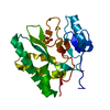
- PDB-8xig: The crystal structure of the AEP domain of MPXV E5 -
+
Open data
ID or keywords:
Loading...
-
Basic information
Entry
Database: PDB / ID: 8xig
Title
The crystal structure of the AEP domain of MPXV E5
Components
Uncoating factor OPG117
Keywords
REPLICATION / primase
Function / homology
Function and homology information
helicase activity / Hydrolases; Acting on acid anhydrides; Acting on acid anhydrides to facilitate cellular and subcellular movement / host cell cytoplasm / hydrolase activity / ATP binding Similarity search - Function
DNA primase/nucleoside triphosphatase, C-terminal / Poxvirus D5 protein-like / : / Bacteriophage/plasmid primase, P4, C-terminal / D5 N terminal like / Helicase, superfamily 3, DNA virus / Superfamily 3 helicase of DNA viruses domain profile. / P-loop containing nucleoside triphosphate hydrolase Similarity search - Domain/homology
National Natural Science Foundation of China (NSFC)
China
Citation
Journal: Cell Discov / Year: 2024 Title: Structural and functional insights into the helicase protein E5 of Mpox virus. Authors: Weizhen Zhang / Yusong Liu / Mengquan Yang / Jie Yang / Zhiwei Shao / Yanqing Gao / Xinran Jiang / Ruixue Cui / Yixi Zhang / Xin Zhao / Qiyuan Shao / Chulei Cao / Huili Li / Linxi Li / Hehua ...Authors: Weizhen Zhang / Yusong Liu / Mengquan Yang / Jie Yang / Zhiwei Shao / Yanqing Gao / Xinran Jiang / Ruixue Cui / Yixi Zhang / Xin Zhao / Qiyuan Shao / Chulei Cao / Huili Li / Linxi Li / Hehua Liu / Haishan Gao / Jianhua Gan / Abstract: Mpox virus (MPXV) can cause mpox in humans. Due to its quick and wide spread in the past two years, mpox has turned into a significant public health concern. Helicase E5 is a multi-domain protein; ...Mpox virus (MPXV) can cause mpox in humans. Due to its quick and wide spread in the past two years, mpox has turned into a significant public health concern. Helicase E5 is a multi-domain protein; its primer synthesis and DNA unwinding activity are required for genome uncoating and DNA replication of MPXV. However, the in vitro DNA unwinding activity has never been demonstrated. Here, we report the structural and biochemical studies of MPXV E5, showing that the full-length protein adopts an auto-inhibited conformation. Truncation of the N-terminus can recover the in vitro unwinding activity of E5 towards the forked DNA. Further structural analysis reveals that MPXV E5 shares a conserved mechanism in DNA unwinding and primer synthesis with the homologous proteins. These findings not only advance our understanding on the function of MPXV E5, but also provide a solid basis for the development of anti-poxvirus drugs.
Mass: 25940.324 Da / Num. of mol.: 1 Source method: isolated from a genetically manipulated source Source: (gene. exp.) Monkeypox virus / Gene: OPG117, MPXVgp100 / Production host: Escherichia coli (E. coli) References: UniProt: A0A7H0DN89, Hydrolases; Acting on acid anhydrides; Acting on acid anhydrides to facilitate cellular and subcellular movement
In the structure databanks used in Yorodumi, some data are registered as the other names, "COVID-19 virus" and "2019-nCoV". Here are the details of the virus and the list of structure data.
Jan 31, 2019. EMDB accession codes are about to change! (news from PDBe EMDB page)
EMDB accession codes are about to change! (news from PDBe EMDB page)
The allocation of 4 digits for EMDB accession codes will soon come to an end. Whilst these codes will remain in use, new EMDB accession codes will include an additional digit and will expand incrementally as the available range of codes is exhausted. The current 4-digit format prefixed with “EMD-” (i.e. EMD-XXXX) will advance to a 5-digit format (i.e. EMD-XXXXX), and so on. It is currently estimated that the 4-digit codes will be depleted around Spring 2019, at which point the 5-digit format will come into force.
The EM Navigator/Yorodumi systems omit the EMD- prefix.
Related info.:Q: What is EMD? / ID/Accession-code notation in Yorodumi/EM Navigator
Yorodumi is a browser for structure data from EMDB, PDB, SASBDB, etc.
This page is also the successor to EM Navigator detail page, and also detail information page/front-end page for Omokage search.
The word "yorodu" (or yorozu) is an old Japanese word meaning "ten thousand". "mi" (miru) is to see.
Related info.:EMDB / PDB / SASBDB / Comparison of 3 databanks / Yorodumi Search / Aug 31, 2016. New EM Navigator & Yorodumi / Yorodumi Papers / Jmol/JSmol / Function and homology information / Changes in new EM Navigator and Yorodumi
 Movie
Movie Controller


 Open data
 Basic information
 Components
 Keywords
 Function and homology information
 Monkeypox virus
 X-RAY DIFFRACTION /
  Authors
 China, 1items
  Citation
 Structure visualization
 Downloads & links
 Other downloads


 PDBj
PDBj

 Assembly

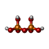
 Mass: 177.975 Da / Num. of mol.: 1 / Source method: obtained synthetically / Formula: H4O7P2 / Feature type: SUBJECT OF INVESTIGATION
 Mass: 24.305 Da / Num. of mol.: 2 / Source method: obtained synthetically / Formula: Mg / Feature type: SUBJECT OF INVESTIGATION
 Mass: 18.015 Da / Num. of mol.: 358 / Source method: isolated from a natural source / Formula: H2O
 Sample preparation
 Processing