 Movie
Movie Controller

[English] 日本語
 Yorodumi
- PDB-8xfe: Cryo-EM structure of defence-associated sirtuin 2 (DSR2) H171A pr... -
+ Open data

- Basic information
| Entry | Database: PDB / ID: 8xfe | ||||||
|---|---|---|---|---|---|---|---|
| Title | Cryo-EM structure of defence-associated sirtuin 2 (DSR2) H171A protein in complex with DSR anti-defence 1(DSAD1) | ||||||
 Components Components |
| ||||||
 Keywords Keywords | CELL INVASION / Cryo-EM / defence-associated sirtuin (DSR)DSR2 / DSR anti-defence 1(DSAD1) / Phage invasion | ||||||
| Biological species |   Phage #D (virus) Phage #D (virus) | ||||||
| Method | ELECTRON MICROSCOPY / single particle reconstruction / cryo EM / Resolution: 2.98 Å | ||||||
 Authors Authors | Li, Y. / Zhang, H. / Zheng, Q. / Wu, Y. / Li, S. | ||||||
| Funding support | 1items
| ||||||
 Citation Citation | Journal: Sci Adv / Year: 2024 Title: Structural insights into activation mechanisms on NADase of the bacterial DSR2 anti-phage defense system. Authors: Hong Zhang / Yu Li / Lanlan Li / Lifei Chen / Chunhua Zhu / Lifang Sun / Panpan Dong / Dingding Jing / Jinbo Yang / Lei Fu / Fangnan Xiao / Ningshao Xia / Shaowei Li / Qingbing Zheng / Yunkun Wu /  Abstract: As a sirtuin (SIR2) family protein, defense-associated sirtuin2 (DSR2) has been demonstrated to participate in bacterial anti-phage resistance via depleting nicotinamide adenine dinucleotide (NAD) of ...As a sirtuin (SIR2) family protein, defense-associated sirtuin2 (DSR2) has been demonstrated to participate in bacterial anti-phage resistance via depleting nicotinamide adenine dinucleotide (NAD) of infected cells, which can be activated by tail tube protein (TTP) and inhibited by DSR anti-defense 1 (DSAD1) of diverse phages. However, the regulating mechanism remains elusive. Here, we determined the cryo-electron microscopy structure of apo DSR2, as well as the respective complex structures with TTP and DSAD1. Structural analyses and biochemical studies reveal that DSR2 forms a tetramer with a SIR2 central core and two distinct conformations. Monomeric TTP preferentially binds to the closed conformation of DSR2, inducing conformational distortions on SIR2 tetramer assembly to activate its NADase activity. DSAD1 combines with the open conformation of DSR2, directly or allosterically inhibiting TTP activation on DSR2 NAD hydrolysis. Our findings decipher the detailed molecule mechanisms for DSR2 NADase activity regulation and lay a foundation for in-depth understanding of the DSR2 anti-phage defense system. | ||||||
| History |
|
- Structure visualization
| Structure viewer | Molecule:  MolmilJmol/JSmol MolmilJmol/JSmol |
|---|
- Downloads & links
-Download
| PDBx/mmCIF format | 8xfe.cif.gz | 479.4 KB | Display | PDBx/mmCIF format |
|---|---|---|---|---|
| PDB format | pdb8xfe.ent.gz | 371.8 KB | Display | PDB format |
| PDBx/mmJSON format | 8xfe.json.gz | Tree view | PDBx/mmJSON format | |
| Others |  Other downloads Other downloads |
-Validation report
| Summary document | 8xfe_validation.pdf.gz | 1.4 MB | Display | wwPDB validaton report |
|---|---|---|---|---|
| Full document | 8xfe_full_validation.pdf.gz | 1.4 MB | Display | |
| Data in XML | 8xfe_validation.xml.gz | 75.1 KB | Display | |
| Data in CIF | 8xfe_validation.cif.gz | 114.3 KB | Display | |
| Arichive directory | https://data.pdbj.org/pub/pdb/validation_reports/xf/8xfeftp://data.pdbj.org/pub/pdb/validation_reports/xf/8xfe | HTTPS FTP |
-Related structure data
| Related structure data | 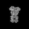 38302MC  8xewC 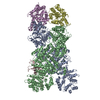 8xffC M: map data used to model this data C: citing same article ( |
|---|


-Links
 PDBj
PDBj- Assembly
| Deposited unit | 
|
|---|---|
| 1 |
|
-Components
| #1: Protein | Mass: 118568.727 Da / Num. of mol.: 4 Source method: isolated from a genetically manipulated source Details: WP_029317421.1 / Source: (gene. exp.) Production host:  Bacteria Latreille et al. 1825 (Bacteria stick insect) Bacteria Latreille et al. 1825 (Bacteria stick insect)#2: Protein | | Mass: 13239.132 Da / Num. of mol.: 1 Source method: isolated from a genetically manipulated source Source: (gene. exp.) Phage #D (virus)Production host: Bacteria Latreille et al. 1825 (Bacteria stick insect)Has protein modification | N | |
|---|
-Experimental details
-Experiment
| Experiment | Method: ELECTRON MICROSCOPY |
|---|---|
| EM experiment | Aggregation state: PARTICLE / 3D reconstruction method: single particle reconstruction |
- Sample preparation
| Component |
| ||||||||||||||||||||||||
|---|---|---|---|---|---|---|---|---|---|---|---|---|---|---|---|---|---|---|---|---|---|---|---|---|---|
| Source (natural) |
| ||||||||||||||||||||||||
| Source (recombinant) | Organism: Bacteria Latreille et al. 1825 (Bacteria stick insect) | ||||||||||||||||||||||||
| Buffer solution | pH: 7.4 | ||||||||||||||||||||||||
| Specimen | Embedding applied: NO / Shadowing applied: NO / Staining applied: NO / Vitrification applied: YES | ||||||||||||||||||||||||
| Vitrification | Cryogen name: ETHANE |
- Electron microscopy imaging
| Experimental equipment |  Model: Tecnai F30 / Image courtesy: FEI Company |
|---|---|
| Microscopy | Model: FEI TECNAI F30 |
| Electron gun | Electron source:  FIELD EMISSION GUN / Accelerating voltage: 300 kV / Illumination mode: FLOOD BEAM FIELD EMISSION GUN / Accelerating voltage: 300 kV / Illumination mode: FLOOD BEAM |
| Electron lens | Mode: BRIGHT FIELD / Nominal defocus max: 1800 nm / Nominal defocus min: 900 nm |
| Image recording | Electron dose: 60 e/Å2 / Film or detector model: GATAN K3 (6k x 4k) |
- Processing
| CTF correction | Type: PHASE FLIPPING AND AMPLITUDE CORRECTION |
|---|---|
| 3D reconstruction | Resolution: 2.98 Å / Resolution method: FSC 0.143 CUT-OFF / Num. of particles: 269385 / Symmetry type: POINT |
