 Movie
Movie Controller

[English] 日本語
 Yorodumi
- PDB-8xb2: Structure of radafaxine-bound state of the human Norepinephrine T... -
+ Open data

- Basic information
| Entry | Database: PDB / ID: 8xb2 | ||||||
|---|---|---|---|---|---|---|---|
| Title | Structure of radafaxine-bound state of the human Norepinephrine Transporter | ||||||
 Components Components | GFP-MBP-solute carrier family 6 member 2,Maltose/maltodextrin-binding periplasmic protein,Sodium-dependent noradrenaline transporter,Maltose/maltodextrin-binding periplasmic protein,Sodium-dependent noradrenaline transporter | ||||||
 Keywords Keywords | TRANSPORT PROTEIN / antidepressant / bupropion | ||||||
| Function / homology |  Function and homology information Function and homology informationneurotransmitter:sodium symporter activity / Defective SLC6A2 causes orthostatic intolerance (OI) / norepinephrine uptake / : / norepinephrine:sodium symporter activity / dopamine:sodium symporter activity / neurotransmitter transmembrane transporter activity / monoamine transmembrane transporter activity / : / SLC-mediated transport of neurotransmitters ...neurotransmitter:sodium symporter activity / Defective SLC6A2 causes orthostatic intolerance (OI) / norepinephrine uptake / : / norepinephrine:sodium symporter activity / dopamine:sodium symporter activity / neurotransmitter transmembrane transporter activity / monoamine transmembrane transporter activity / : / SLC-mediated transport of neurotransmitters / response to pain / neurotransmitter transport / neuronal cell body membrane / detection of maltose stimulus / maltose transport complex / dopamine uptake involved in synaptic transmission / carbohydrate transport / amino acid transport / carbohydrate transmembrane transporter activity / maltose binding / maltose transport / maltodextrin transmembrane transport / alpha-tubulin binding / beta-tubulin binding / ATP-binding cassette (ABC) transporter complex, substrate-binding subunit-containing / ATP-binding cassette (ABC) transporter complex / sodium ion transmembrane transport / cell chemotaxis / neuron cellular homeostasis / outer membrane-bounded periplasmic space / synaptic vesicle membrane / actin binding / presynaptic membrane / chemical synaptic transmission / periplasmic space / response to xenobiotic stimulus / axon / DNA damage response / cell surface / membrane / metal ion binding / plasma membrane Similarity search - Function | ||||||
| Biological species |  Homo sapiens (human) Homo sapiens (human) | ||||||
| Method | ELECTRON MICROSCOPY / single particle reconstruction / cryo EM / Resolution: 3.04 Å | ||||||
 Authors Authors | Wu, J.X. / Ji, W.M. | ||||||
| Funding support |  China, 1items China, 1items
| ||||||
 Citation Citation | Journal: Nature / Year: 2024 Title: Substrate binding and inhibition mechanism of norepinephrine transporter. Authors: Wenming Ji / Anran Miao / Kai Liang / Jiameng Liu / Yuhan Qi / Yue Zhou / Xinli Duan / Jixue Sun / Lipeng Lai / Jing-Xiang Wu / Abstract: Norepinephrine transporter (NET; encoded by SLC6A2) reuptakes the majority of the released noradrenaline back to the presynaptic terminals, thereby affecting the synaptic noradrenaline level. Genetic ...Norepinephrine transporter (NET; encoded by SLC6A2) reuptakes the majority of the released noradrenaline back to the presynaptic terminals, thereby affecting the synaptic noradrenaline level. Genetic mutations and dysregulation of NET are associated with a spectrum of neurological conditions in humans, making NET an important therapeutic target. However, the structure and mechanism of NET remain unclear. Here we provide cryogenic electron microscopy structures of the human NET (hNET) in three functional states-the apo state, and in states bound to the substrate meta-iodobenzylguanidine (MIBG) or the orthosteric inhibitor radafaxine. These structures were captured in an inward-facing conformation, with a tightly sealed extracellular gate and an open intracellular gate. The substrate MIBG binds at the centre of hNET. Radafaxine also occupies the substrate-binding site and might block the structural transition of hNET for inhibition. These structures provide insights into the mechanism of substrate recognition and orthosteric inhibition of hNET. | ||||||
| History |
|
- Structure visualization
| Structure viewer | Molecule: MolmilJmol/JSmol |
|---|
- Downloads & links
-Download
| PDBx/mmCIF format | 8xb2.cif.gz | 125.4 KB | Display | PDBx/mmCIF format |
|---|---|---|---|---|
| PDB format | pdb8xb2.ent.gz | 84.6 KB | Display | PDB format |
| PDBx/mmJSON format | 8xb2.json.gz | Tree view | PDBx/mmJSON format | |
| Others |  Other downloads Other downloads |
-Validation report
| Arichive directory | https://data.pdbj.org/pub/pdb/validation_reports/xb/8xb2ftp://data.pdbj.org/pub/pdb/validation_reports/xb/8xb2 | HTTPS FTP |
|---|
-Related structure data
| Related structure data |  38208MC 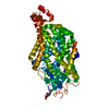 8xb3C 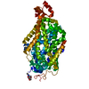 8xb4C M: map data used to model this data C: citing same article ( |
|---|---|
| Similar structure data |

-Links
 PDBj
PDBj


- Assembly
| Deposited unit | 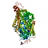
|
|---|---|
| 1 |
|
-Components
| #1: Protein | Mass: 138697.016 Da / Num. of mol.: 1 Source method: isolated from a genetically manipulated source Source: (gene. exp.) Homo sapiens (human), (gene. exp.) Gene: SLC6A2, malE, b4034, JW3994, SLC6A2, NAT1, NET1, SLC6A5 Cell line (production host): HEK293 / Production host: Homo sapiens (human) / References: UniProt: P0AEX9, UniProt: P23975 |
|---|---|
| #2: Sugar | ChemComp-NAG /   Type: D-saccharide, beta linking / Mass: 221.208 Da / Num. of mol.: 1 / Source method: obtained synthetically / Formula: C8H15NO6 Type: D-saccharide, beta linking / Mass: 221.208 Da / Num. of mol.: 1 / Source method: obtained synthetically / Formula: C8H15NO6 |
| #3: Chemical | ChemComp-YNT / Mass: 255.741 Da / Num. of mol.: 1 / Source method: obtained synthetically / Formula: C13H18ClNO2 |
| Has ligand of interest | Y |
| Has protein modification | Y |
-Experimental details
-Experiment
| Experiment | Method: ELECTRON MICROSCOPY |
|---|---|
| EM experiment | Aggregation state: PARTICLE / 3D reconstruction method: single particle reconstruction |
- Sample preparation
| Component | Name: NET / Type: COMPLEX / Entity ID: #1 / Source: RECOMBINANT |
|---|---|
| Source (natural) | Organism: Homo sapiens (human) |
| Source (recombinant) | Organism: Homo sapiens (human) |
| Buffer solution | pH: 7.5 |
| Specimen | Embedding applied: NO / Shadowing applied: NO / Staining applied: NO / Vitrification applied: YES |
| Vitrification | Cryogen name: ETHANE |
- Electron microscopy imaging
| Experimental equipment |  Model: Titan Krios / Image courtesy: FEI Company |
|---|---|
| Microscopy | Model: FEI TITAN KRIOS |
| Electron gun | Electron source:  FIELD EMISSION GUN / Accelerating voltage: 300 kV / Illumination mode: FLOOD BEAM FIELD EMISSION GUN / Accelerating voltage: 300 kV / Illumination mode: FLOOD BEAM |
| Electron lens | Mode: BRIGHT FIELD / Calibrated magnification: 64000 X / Nominal defocus max: 2000 nm / Nominal defocus min: 1000 nm |
| Image recording | Electron dose: 50 e/Å2 / Film or detector model: GATAN K3 BIOQUANTUM (6k x 4k) |
- Processing
| EM software | Name: PHENIX / Version: 1.16_3549: / Category: model refinement | ||||||||||||||||||||||||
|---|---|---|---|---|---|---|---|---|---|---|---|---|---|---|---|---|---|---|---|---|---|---|---|---|---|
| CTF correction | Type: NONE | ||||||||||||||||||||||||
| 3D reconstruction | Resolution: 3.04 Å / Resolution method: FSC 0.143 CUT-OFF / Num. of particles: 146222 / Symmetry type: POINT | ||||||||||||||||||||||||
| Refine LS restraints |
|
