 Movie
Movie Controller

[English] 日本語
 Yorodumi
- PDB-8x8s: Crystal structure of Cypovirus Polyhedra mutant fused with c-Myc ... -
+ Open data

- Basic information
| Entry | Database: PDB / ID: 8x8s | ||||||
|---|---|---|---|---|---|---|---|
| Title | Crystal structure of Cypovirus Polyhedra mutant fused with c-Myc fragment | ||||||
 Components Components | Polyhedrin,Myc proto-oncogene protein | ||||||
 Keywords Keywords | VIRAL PROTEIN / Polyhedra | ||||||
| Function / homology |  Function and homology information Function and homology informationpositive regulation of metanephric cap mesenchymal cell proliferation / positive regulation of acinar cell proliferation / acinar cell proliferation / SCF ubiquitin ligase complex binding / NK T cell proliferation / Myc-Max complex / viral occlusion body / regulation of somatic stem cell population maintenance / regulation of cell cycle process / RNA polymerase II transcription repressor complex ...positive regulation of metanephric cap mesenchymal cell proliferation / positive regulation of acinar cell proliferation / acinar cell proliferation / SCF ubiquitin ligase complex binding / NK T cell proliferation / Myc-Max complex / viral occlusion body / regulation of somatic stem cell population maintenance / regulation of cell cycle process / RNA polymerase II transcription repressor complex / Binding of TCF/LEF:CTNNB1 to target gene promoters / cellular response to interferon-alpha / positive regulation of B cell apoptotic process / RUNX3 regulates WNT signaling / TFAP2 (AP-2) family regulates transcription of cell cycle factors / myotube differentiation / negative regulation of cell division / negative regulation of transcription initiation by RNA polymerase II / Regulation of CDH1 mRNA translation by microRNAs / negative regulation of monocyte differentiation / detection of mechanical stimulus involved in sensory perception of sound / response to growth factor / B cell apoptotic process / response to alkaloid / transcription regulator activator activity / Transcription of E2F targets under negative control by DREAM complex / fibroblast apoptotic process / negative regulation of stress-activated MAPK cascade / Regulation of NFE2L2 gene expression / positive regulation of mesenchymal cell proliferation / protein-DNA complex disassembly / skeletal system morphogenesis / regulation of telomere maintenance / branching involved in ureteric bud morphogenesis / Signaling by ALK / middle ear morphogenesis / rRNA metabolic process / negative regulation of gene expression via chromosomal CpG island methylation / E-box binding / pigmentation / positive regulation of telomere maintenance / skeletal muscle cell differentiation / chromosome organization / positive regulation of transcription initiation by RNA polymerase II / positive regulation of intrinsic apoptotic signaling pathway by p53 class mediator / Cyclin E associated events during G1/S transition / core promoter sequence-specific DNA binding / Cyclin A:Cdk2-associated events at S phase entry / negative regulation of fibroblast proliferation / ERK1 and ERK2 cascade / positive regulation of epithelial cell proliferation / transcription coregulator binding / euchromatin / G1/S transition of mitotic cell cycle / SMAD2/SMAD3:SMAD4 heterotrimer regulates transcription / protein processing / positive regulation of miRNA transcription / DNA-binding transcription repressor activity, RNA polymerase II-specific / MAPK6/MAPK4 signaling / NOTCH1 Intracellular Domain Regulates Transcription / cellular response to xenobiotic stimulus / positive regulation of fibroblast proliferation / spindle / intrinsic apoptotic signaling pathway in response to DNA damage / Constitutive Signaling by NOTCH1 PEST Domain Mutants / Constitutive Signaling by NOTCH1 HD+PEST Domain Mutants / Wnt signaling pathway / Transcriptional regulation of granulopoiesis / cellular response to UV / MAPK cascade / regulation of gene expression / Interleukin-4 and Interleukin-13 signaling / DNA-binding transcription activator activity, RNA polymerase II-specific / cellular response to hypoxia / transcription by RNA polymerase II / Estrogen-dependent gene expression / DNA-binding transcription factor binding / host cell cytoplasm / intracellular iron ion homeostasis / DNA-binding transcription factor activity, RNA polymerase II-specific / protein dimerization activity / Ub-specific processing proteases / nuclear body / RNA polymerase II cis-regulatory region sequence-specific DNA binding / chromatin remodeling / response to xenobiotic stimulus / axon / positive regulation of cell population proliferation / DNA damage response / ubiquitin protein ligase binding / positive regulation of gene expression / regulation of transcription by RNA polymerase II / negative regulation of apoptotic process / positive regulation of DNA-templated transcription / chromatin / protein-containing complex binding / perinuclear region of cytoplasm / nucleolus / negative regulation of transcription by RNA polymerase II / positive regulation of transcription by RNA polymerase II Similarity search - Function | ||||||
| Biological species |   Bombyx mori cypovirus 1 Bombyx mori cypovirus 1 Homo sapiens (human) Homo sapiens (human) | ||||||
| Method |  X-RAY DIFFRACTION / SYNCHROTRON / MOLECULAR REPLACEMENT / Resolution: 2.04 Å X-RAY DIFFRACTION / SYNCHROTRON / MOLECULAR REPLACEMENT / Resolution: 2.04 Å | ||||||
 Authors Authors | Kojima, M. / Ueno, T. / Abe, S. / Hirata, K. | ||||||
| Funding support |  Japan, 1items Japan, 1items
| ||||||
 Citation Citation | Journal: Proc.Natl.Acad.Sci.USA / Year: 2024 Title: High-throughput structure determination of an intrinsically disordered protein using cell-free protein crystallization. Authors: Kojima, M. / Abe, S. / Furuta, T. / Hirata, K. / Yao, X. / Kobayashi, A. / Kobayashi, R. / Ueno, T. | ||||||
| History |
|
- Structure visualization
| Structure viewer | Molecule: MolmilJmol/JSmol |
|---|
- Downloads & links
-Download
| PDBx/mmCIF format | 8x8s.cif.gz | 59.2 KB | Display | PDBx/mmCIF format |
|---|---|---|---|---|
| PDB format | pdb8x8s.ent.gz | 34 KB | Display | PDB format |
| PDBx/mmJSON format | 8x8s.json.gz | Tree view | PDBx/mmJSON format | |
| Others |  Other downloads Other downloads |
-Validation report
| Arichive directory | https://data.pdbj.org/pub/pdb/validation_reports/x8/8x8sftp://data.pdbj.org/pub/pdb/validation_reports/x8/8x8s | HTTPS FTP |
|---|
-Related structure data


-Links
 PDBj
PDBj


- Assembly
| Deposited unit | 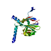
| ||||||||||||
|---|---|---|---|---|---|---|---|---|---|---|---|---|---|
| 1 | 
| ||||||||||||
| Unit cell |
|
-Components
| #1: Protein | Mass: 28301.340 Da / Num. of mol.: 1 / Mutation: R151K Source method: isolated from a genetically manipulated source Details: cell-free synthesis Source: (gene. exp.) Bombyx mori cypovirus 1, (gene. exp.) Homo sapiens (human)Gene: MYC, BHLHE39 / Production host:  |
|---|---|
| #2: Water | ChemComp-HOH /  Mass: 18.015 Da / Num. of mol.: 23 / Source method: isolated from a natural source / Formula: H2O Mass: 18.015 Da / Num. of mol.: 23 / Source method: isolated from a natural source / Formula: H2O |
-Experimental details
-Experiment
| Experiment | Method: X-RAY DIFFRACTION / Number of used crystals: 1 |
|---|
- Sample preparation
| Crystal | Density Matthews: 1.78 Å3/Da / Density % sol: 30.9 % |
|---|---|
| Crystal grow | Temperature: 293 K / Method: small tubes / Details: cell-free crystallization |
-Data collection
| Diffraction | Mean temperature: 100 K / Serial crystal experiment: N |
|---|---|
| Diffraction source | Source: SYNCHROTRON / Site: SPring-8 / Beamline: BL32XU / Wavelength: 1 Å |
| Detector | Type: DECTRIS PILATUS3 6M / Detector: PIXEL / Date: Nov 17, 2022 |
| Radiation | Protocol: SINGLE WAVELENGTH / Monochromatic (M) / Laue (L): M / Scattering type: x-ray |
| Radiation wavelength | Wavelength: 1 Å / Relative weight: 1 |
| Reflection | Resolution: 2.04→50 Å / Num. obs: 12981 / % possible obs: 99.9 % / Redundancy: 58 % / Biso Wilson estimate: 33.27 Å2 / CC1/2: 0.999 / Net I/σ(I): 19.4 |
| Reflection shell | Resolution: 2.04→2.11 Å / Num. unique obs: 2027 / CC1/2: 0.658 |
- Processing
| Software |
| ||||||||||||||||||||||||||||||||||||||||||||||||||||||||||||||||||||||
|---|---|---|---|---|---|---|---|---|---|---|---|---|---|---|---|---|---|---|---|---|---|---|---|---|---|---|---|---|---|---|---|---|---|---|---|---|---|---|---|---|---|---|---|---|---|---|---|---|---|---|---|---|---|---|---|---|---|---|---|---|---|---|---|---|---|---|---|---|---|---|---|
| Refinement | Method to determine structure: MOLECULAR REPLACEMENT / Resolution: 2.04→43.49 Å / SU ML: 0.3037 / Cross valid method: FREE R-VALUE / σ(F): 1.34 / Phase error: 32.1416 Stereochemistry target values: GeoStd + Monomer Library + CDL v1.2
| ||||||||||||||||||||||||||||||||||||||||||||||||||||||||||||||||||||||
| Solvent computation | Shrinkage radii: 0.9 Å / VDW probe radii: 1.11 Å / Solvent model: FLAT BULK SOLVENT MODEL | ||||||||||||||||||||||||||||||||||||||||||||||||||||||||||||||||||||||
| Displacement parameters | Biso mean: 34.84 Å2 | ||||||||||||||||||||||||||||||||||||||||||||||||||||||||||||||||||||||
| Refinement step | Cycle: LAST / Resolution: 2.04→43.49 Å
| ||||||||||||||||||||||||||||||||||||||||||||||||||||||||||||||||||||||
| Refine LS restraints |
| ||||||||||||||||||||||||||||||||||||||||||||||||||||||||||||||||||||||
| LS refinement shell |
|
