- PDB-8x72: The Crystal Structure of PLK1 from Biortus. -
+
データを開く
IDまたはキーワード:
読み込み中...
-
基本情報
登録情報
データベース: PDB / ID: 8x72
タイトル
The Crystal Structure of PLK1 from Biortus.
要素
Serine/threonine-protein kinase PLK1
キーワード
TRANSFERASE / Kinase Cell cycle
機能・相同性
機能・相同性情報
positive regulation of mitotic nuclear envelope disassembly / Mitotic Telophase/Cytokinesis / regulation of protein localization to cell cortex / Mitotic Metaphase/Anaphase Transition / synaptonemal complex disassembly / Activation of NIMA Kinases NEK9, NEK6, NEK7 / polo kinase / Phosphorylation of Emi1 / homologous chromosome segregation / mitotic nuclear membrane disassembly ...positive regulation of mitotic nuclear envelope disassembly / Mitotic Telophase/Cytokinesis / regulation of protein localization to cell cortex / Mitotic Metaphase/Anaphase Transition / synaptonemal complex disassembly / Activation of NIMA Kinases NEK9, NEK6, NEK7 / polo kinase / Phosphorylation of Emi1 / homologous chromosome segregation / mitotic nuclear membrane disassembly / metaphase/anaphase transition of mitotic cell cycle / female meiosis chromosome segregation / synaptonemal complex / Phosphorylation of the APC/C / anaphase-promoting complex binding / astral microtubule organization / Golgi inheritance / outer kinetochore / mitotic cleavage furrow formation / microtubule bundle formation / mitotic chromosome condensation / double-strand break repair via alternative nonhomologous end joining / Polo-like kinase mediated events / regulation of mitotic spindle assembly / Golgi Cisternae Pericentriolar Stack Reorganization / positive regulation of mitotic metaphase/anaphase transition / centrosome cycle / positive regulation of ubiquitin-dependent protein catabolic process / regulation of mitotic metaphase/anaphase transition / sister chromatid cohesion / regulation of mitotic cell cycle phase transition / mitotic spindle assembly checkpoint signaling / mitotic spindle pole / spindle midzone / mitotic G2 DNA damage checkpoint signaling / regulation of anaphase-promoting complex-dependent catabolic process / mitotic cytokinesis / mitotic sister chromatid segregation / negative regulation of double-strand break repair via homologous recombination / Regulation of MITF-M-dependent genes involved in cell cycle and proliferation / Cyclin A/B1/B2 associated events during G2/M transition / protein localization to chromatin / Amplification of signal from unattached kinetochores via a MAD2 inhibitory signal / Loss of Nlp from mitotic centrosomes / Loss of proteins required for interphase microtubule organization from the centrosome / Recruitment of mitotic centrosome proteins and complexes / Recruitment of NuMA to mitotic centrosomes / Anchoring of the basal body to the plasma membrane / Mitotic Prometaphase / EML4 and NUDC in mitotic spindle formation / regulation of mitotic cell cycle / AURKA Activation by TPX2 / Resolution of Sister Chromatid Cohesion / Condensation of Prophase Chromosomes / mitotic spindle organization / regulation of cytokinesis / establishment of protein localization / centriole / RHO GTPases Activate Formins / APC/C:Cdh1 mediated degradation of Cdc20 and other APC/C:Cdh1 targeted proteins in late mitosis/early G1 / kinetochore / positive regulation of protein localization to nucleus / G2/M transition of mitotic cell cycle / spindle / centriolar satellite / spindle pole / The role of GTSE1 in G2/M progression after G2 checkpoint / Separation of Sister Chromatids / Regulation of PLK1 Activity at G2/M Transition / double-strand break repair / mitotic cell cycle / positive regulation of proteasomal ubiquitin-dependent protein catabolic process / microtubule cytoskeleton / midbody / microtubule binding / protein phosphorylation / protein kinase activity / regulation of cell cycle / protein serine kinase activity / protein serine/threonine kinase activity / centrosome / protein kinase binding / negative regulation of apoptotic process / chromatin / magnesium ion binding / negative regulation of transcription by RNA polymerase II / nucleoplasm / ATP binding / identical protein binding / nucleus / cytoplasm / cytosol 類似検索 - 分子機能
Polo-like kinase 1, catalytic domain / Second polo-box domain / First polo-box domain / POLO box domain superfamily / POLO box duplicated region / POLO box domain / POLO box domain profile. / Serine/threonine-protein kinase, active site / Serine/Threonine protein kinases active-site signature. / Protein kinase domain ...Polo-like kinase 1, catalytic domain / Second polo-box domain / First polo-box domain / POLO box domain superfamily / POLO box duplicated region / POLO box domain / POLO box domain profile. / Serine/threonine-protein kinase, active site / Serine/Threonine protein kinases active-site signature. / Protein kinase domain / Serine/Threonine protein kinases, catalytic domain / Protein kinase, ATP binding site / Protein kinases ATP-binding region signature. / Protein kinase domain profile. / Protein kinase domain / Protein kinase-like domain superfamily 類似検索 - ドメイン・相同性
 ムービー
ムービー コントローラー


 データを開く
 基本情報
基本情報 要素
要素 キーワード
キーワード 機能・相同性情報
 Homo sapiens (ヒト)
 X線回折 /
  データ登録者
 引用
引用 構造の表示
構造の表示 ダウンロードとリンク
 その他のダウンロード
 PDBj
PDBj


 集合体
集合体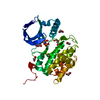


 分子量: 506.196 Da / 分子数: 1 / 由来タイプ: 合成 / 式: C10H17N6O12P3
 分子量: 65.409 Da / 分子数: 1 / 由来タイプ: 合成 / 式: Zn
 分子量: 59.044 Da / 分子数: 2 / 由来タイプ: 合成 / 式: C2H3O2
 分子量: 106.120 Da / 分子数: 2 / 由来タイプ: 合成 / 式: C4H10O3
 分子量: 62.068 Da / 分子数: 3 / 由来タイプ: 合成 / 式: C2H6O2
 試料調製
試料調製 / ビームライン: MX2 / 波長: 0.95365 Å
 解析
解析