 Movie
Movie Controller


+ Open data

- Basic information
| Entry | Database: PDB / ID: 8x70 | ||||||
|---|---|---|---|---|---|---|---|
| Title | The Crystal Structure of IFI16 from Biortus. | ||||||
 Components Components | Gamma-interferon-inducible protein 16 | ||||||
 Keywords Keywords | PROTEIN BINDING / Activator / DNA-binding / Apoptosis | ||||||
| Function / homology |  Function and homology information Function and homology informationnegative regulation of AIM2 inflammasome complex assembly / negative regulation of DNA binding / myeloid cell differentiation / STING mediated induction of host immune responses / IRF3-mediated induction of type I IFN / negative regulation of viral genome replication / transcription factor binding / negative regulation of gene expression, epigenetic / intrinsic apoptotic signaling pathway by p53 class mediator / intrinsic apoptotic signaling pathway in response to DNA damage by p53 class mediator ...negative regulation of AIM2 inflammasome complex assembly / negative regulation of DNA binding / myeloid cell differentiation / STING mediated induction of host immune responses / IRF3-mediated induction of type I IFN / negative regulation of viral genome replication / transcription factor binding / negative regulation of gene expression, epigenetic / intrinsic apoptotic signaling pathway by p53 class mediator / intrinsic apoptotic signaling pathway in response to DNA damage by p53 class mediator / monocyte differentiation / cellular response to glucose starvation / cellular response to interferon-beta / activation of innate immune response / negative regulation of innate immune response / positive regulation of cytokine production / positive regulation of interleukin-1 beta production / regulation of autophagy / cellular response to ionizing radiation / autophagy / regulation of inflammatory response / double-stranded DNA binding / defense response to virus / nuclear speck / inflammatory response / innate immune response / negative regulation of DNA-templated transcription / nucleolus / negative regulation of transcription by RNA polymerase II / positive regulation of transcription by RNA polymerase II / RNA binding / nucleoplasm / membrane / identical protein binding / nucleus / cytoplasm / cytosol Similarity search - Function | ||||||
| Biological species |  Homo sapiens (human) Homo sapiens (human) | ||||||
| Method |  X-RAY DIFFRACTION / SYNCHROTRON / MOLECULAR REPLACEMENT / Resolution: 1.7 Å X-RAY DIFFRACTION / SYNCHROTRON / MOLECULAR REPLACEMENT / Resolution: 1.7 Å | ||||||
 Authors Authors | Wang, F. / Cheng, W. / Lv, Z. / Meng, Q. / Wang, J. | ||||||
| Funding support | 1items
| ||||||
 Citation Citation | Journal: To Be Published Title: The Crystal Structure of IFI16 from Biortus. Authors: Wang, F. / Cheng, W. / Lv, Z. / Meng, Q. / Wang, J. | ||||||
| History |
|
- Structure visualization
| Structure viewer | Molecule: MolmilJmol/JSmol |
|---|
- Downloads & links
-Download
| PDBx/mmCIF format | 8x70.cif.gz | 191.1 KB | Display | PDBx/mmCIF format |
|---|---|---|---|---|
| PDB format | pdb8x70.ent.gz | 144.1 KB | Display | PDB format |
| PDBx/mmJSON format | 8x70.json.gz | Tree view | PDBx/mmJSON format | |
| Others |  Other downloads Other downloads |
-Validation report
| Arichive directory | https://data.pdbj.org/pub/pdb/validation_reports/x7/8x70ftp://data.pdbj.org/pub/pdb/validation_reports/x7/8x70 | HTTPS FTP |
|---|
-Related structure data
| Similar structure data |
|---|
-Links
 PDBj
PDBj


- Assembly
| Deposited unit | 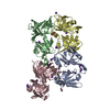
| ||||||||
|---|---|---|---|---|---|---|---|---|---|
| 1 | 
| ||||||||
| 2 | 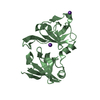
| ||||||||
| 3 | 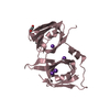
| ||||||||
| 4 | 
| ||||||||
| Unit cell |
|
-Components
-Protein , 1 types, 4 molecules ABCD
| #1: Protein | Mass: 23527.326 Da / Num. of mol.: 4 Source method: isolated from a genetically manipulated source Source: (gene. exp.) Homo sapiens (human) / Gene: IFI16 / Production host:  |
|---|
-Non-polymers , 5 types, 813 molecules 


| #2: Chemical | ChemComp-BR /  Mass: 79.904 Da / Num. of mol.: 1 / Source method: obtained synthetically / Formula: Br Mass: 79.904 Da / Num. of mol.: 1 / Source method: obtained synthetically / Formula: Br | ||||||
|---|---|---|---|---|---|---|---|
| #3: Chemical | ChemComp-EDO /  Mass: 62.068 Da / Num. of mol.: 5 / Source method: obtained synthetically / Formula: C2H6O2 Mass: 62.068 Da / Num. of mol.: 5 / Source method: obtained synthetically / Formula: C2H6O2#4: Chemical | ChemComp-K /  Mass: 39.098 Da / Num. of mol.: 14 / Source method: obtained synthetically / Formula: K Mass: 39.098 Da / Num. of mol.: 14 / Source method: obtained synthetically / Formula: K#5: Chemical | ChemComp-PGE / |  Mass: 150.173 Da / Num. of mol.: 1 / Source method: obtained synthetically / Formula: C6H14O4 Mass: 150.173 Da / Num. of mol.: 1 / Source method: obtained synthetically / Formula: C6H14O4#6: Water | ChemComp-HOH / | Mass: 18.015 Da / Num. of mol.: 792 / Source method: isolated from a natural source / Formula: H2O |
-Details
| Has ligand of interest | N |
|---|
-Experimental details
-Experiment
| Experiment | Method: X-RAY DIFFRACTION / Number of used crystals: 1 |
|---|
- Sample preparation
| Crystal | Density Matthews: 2.17 Å3/Da / Density % sol: 43.22 % |
|---|---|
| Crystal grow | Temperature: 293 K / Method: vapor diffusion, sitting drop / Details: 0.15M K bromide, 30% w/v PEG 2000 MME |
-Data collection
| Diffraction | Mean temperature: 100 K / Serial crystal experiment: N |
|---|---|
| Diffraction source | Source: SYNCHROTRON / Site: CLSI  / Beamline: 08ID-1 / Wavelength: 1.18075 Å / Beamline: 08ID-1 / Wavelength: 1.18075 Å |
| Detector | Type: DECTRIS EIGER X 9M / Detector: PIXEL / Date: Sep 24, 2021 |
| Radiation | Protocol: SINGLE WAVELENGTH / Monochromatic (M) / Laue (L): M / Scattering type: x-ray |
| Radiation wavelength | Wavelength: 1.18075 Å / Relative weight: 1 |
| Reflection | Resolution: 1.7→46.523 Å / Num. obs: 83684 / % possible obs: 99.8 % / Redundancy: 6.6 % / Rmerge(I) obs: 0.072 / Net I/σ(I): 14.8 |
| Reflection shell | Resolution: 1.7→1.73 Å / Rmerge(I) obs: 0.798 / Num. unique obs: 4406 |
- Processing
| Software |
| ||||||||||||||||||||||||||||||||||||||||||||||||||||||||||||||||||||||||||||||||||||||||||||||||||||||||||||||||||||||||||||||||||||||||||||||||||||||||||||||||
|---|---|---|---|---|---|---|---|---|---|---|---|---|---|---|---|---|---|---|---|---|---|---|---|---|---|---|---|---|---|---|---|---|---|---|---|---|---|---|---|---|---|---|---|---|---|---|---|---|---|---|---|---|---|---|---|---|---|---|---|---|---|---|---|---|---|---|---|---|---|---|---|---|---|---|---|---|---|---|---|---|---|---|---|---|---|---|---|---|---|---|---|---|---|---|---|---|---|---|---|---|---|---|---|---|---|---|---|---|---|---|---|---|---|---|---|---|---|---|---|---|---|---|---|---|---|---|---|---|---|---|---|---|---|---|---|---|---|---|---|---|---|---|---|---|---|---|---|---|---|---|---|---|---|---|---|---|---|---|---|---|---|
| Refinement | Method to determine structure: MOLECULAR REPLACEMENT / Resolution: 1.7→46.523 Å / Cor.coef. Fo:Fc: 0.957 / Cor.coef. Fo:Fc free: 0.938 / WRfactor Rfree: 0.256 / WRfactor Rwork: 0.206 / SU B: 2.923 / SU ML: 0.095 / Average fsc free: 0.8906 / Average fsc work: 0.9098 / Cross valid method: FREE R-VALUE / ESU R: 0.129 / ESU R Free: 0.127 Details: Hydrogens have been added in their riding positions
| ||||||||||||||||||||||||||||||||||||||||||||||||||||||||||||||||||||||||||||||||||||||||||||||||||||||||||||||||||||||||||||||||||||||||||||||||||||||||||||||||
| Solvent computation | Ion probe radii: 0.8 Å / Shrinkage radii: 0.8 Å / VDW probe radii: 1.2 Å / Solvent model: MASK BULK SOLVENT | ||||||||||||||||||||||||||||||||||||||||||||||||||||||||||||||||||||||||||||||||||||||||||||||||||||||||||||||||||||||||||||||||||||||||||||||||||||||||||||||||
| Displacement parameters | Biso mean: 27.626 Å2
| ||||||||||||||||||||||||||||||||||||||||||||||||||||||||||||||||||||||||||||||||||||||||||||||||||||||||||||||||||||||||||||||||||||||||||||||||||||||||||||||||
| Refinement step | Cycle: LAST / Resolution: 1.7→46.523 Å
| ||||||||||||||||||||||||||||||||||||||||||||||||||||||||||||||||||||||||||||||||||||||||||||||||||||||||||||||||||||||||||||||||||||||||||||||||||||||||||||||||
| Refine LS restraints |
| ||||||||||||||||||||||||||||||||||||||||||||||||||||||||||||||||||||||||||||||||||||||||||||||||||||||||||||||||||||||||||||||||||||||||||||||||||||||||||||||||
| LS refinement shell |
|
