 Movie
Movie Controller

[English] 日本語
 Yorodumi
- PDB-8x5y: CryoEM structure of the histamine H1 receptor-BRIL/Anti BRIL Fab ... -
+ Open data

- Basic information
| Entry | Database: PDB / ID: 8x5y | |||||||||||||||||||||||||||||||||
|---|---|---|---|---|---|---|---|---|---|---|---|---|---|---|---|---|---|---|---|---|---|---|---|---|---|---|---|---|---|---|---|---|---|---|
| Title | CryoEM structure of the histamine H1 receptor-BRIL/Anti BRIL Fab complex with astemizole | |||||||||||||||||||||||||||||||||
 Components Components | Histamine H1 receptor,Soluble cytochrome b562 | |||||||||||||||||||||||||||||||||
 Keywords Keywords | MEMBRANE PROTEIN / GPCR | |||||||||||||||||||||||||||||||||
| Function / homology |  Function and homology information Function and homology informationHistamine receptors / histamine receptor activity / regulation of vascular permeability / cellular response to histamine / positive regulation of vasoconstriction / G protein-coupled receptor signaling pathway, coupled to cyclic nucleotide second messenger / electron transport chain / visual learning / regulation of synaptic plasticity / memory ...Histamine receptors / histamine receptor activity / regulation of vascular permeability / cellular response to histamine / positive regulation of vasoconstriction / G protein-coupled receptor signaling pathway, coupled to cyclic nucleotide second messenger / electron transport chain / visual learning / regulation of synaptic plasticity / memory / phospholipase C-activating G protein-coupled receptor signaling pathway / G alpha (q) signalling events / chemical synaptic transmission / periplasmic space / electron transfer activity / iron ion binding / G protein-coupled receptor signaling pathway / inflammatory response / heme binding / synapse / dendrite / plasma membrane / cytosol Similarity search - Function | |||||||||||||||||||||||||||||||||
| Biological species |  Homo sapiens (human) Homo sapiens (human) | |||||||||||||||||||||||||||||||||
| Method | ELECTRON MICROSCOPY / single particle reconstruction / cryo EM / Resolution: 3 Å | |||||||||||||||||||||||||||||||||
 Authors Authors | Wang, D.D. / Guo, Q. / Tao, Y.Y. | |||||||||||||||||||||||||||||||||
| Funding support |  China, 1items China, 1items
| |||||||||||||||||||||||||||||||||
 Citation Citation | Journal: Nat Commun / Year: 2024 Title: Molecular mechanism of antihistamines recognition and regulation of the histamine H receptor. Authors: Dandan Wang / Qiong Guo / Zhangsong Wu / Ming Li / Binbin He / Yang Du / Kaiming Zhang / Yuyong Tao / Abstract: Histamine receptors are a group of G protein-coupled receptors (GPCRs) that play important roles in various physiological and pathophysiological conditions. Antihistamines that target the histamine H ...Histamine receptors are a group of G protein-coupled receptors (GPCRs) that play important roles in various physiological and pathophysiological conditions. Antihistamines that target the histamine H receptor (HR) have been widely used to relieve the symptoms of allergy and inflammation. Here, to uncover the details of the regulation of HR by the known second-generation antihistamines, thereby providing clues for the rational design of newer antihistamines, we determine the cryo-EM structure of HR in the apo form and bound to different antihistamines. In addition to the deep hydrophobic cavity, we identify a secondary ligand-binding site in HR, which potentially may support the introduction of new derivative groups to generate newer antihistamines. Furthermore, these structures show that antihistamines exert inverse regulation by utilizing a shared phenyl group that inserts into the deep cavity and block the movement of the toggle switch residue W428. Together, these results enrich our understanding of GPCR modulation and facilitate the structure-based design of novel antihistamines. | |||||||||||||||||||||||||||||||||
| History |
|
- Structure visualization
| Structure viewer | Molecule: MolmilJmol/JSmol |
|---|
- Downloads & links
-Download
| PDBx/mmCIF format | 8x5y.cif.gz | 68.2 KB | Display | PDBx/mmCIF format |
|---|---|---|---|---|
| PDB format | pdb8x5y.ent.gz | 46.1 KB | Display | PDB format |
| PDBx/mmJSON format | 8x5y.json.gz | Tree view | PDBx/mmJSON format | |
| Others |  Other downloads Other downloads |
-Validation report
| Arichive directory | https://data.pdbj.org/pub/pdb/validation_reports/x5/8x5yftp://data.pdbj.org/pub/pdb/validation_reports/x5/8x5y | HTTPS FTP |
|---|
-Related structure data
| Related structure data |  38075MC  8x5xC 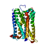 8x63C 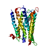 8x64C M: map data used to model this data C: citing same article ( |
|---|---|
| Similar structure data |

-Links
 PDBj
PDBj


- Assembly
| Deposited unit | 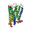
|
|---|---|
| 1 |
|
-Components
| #1: Protein | Mass: 50629.363 Da / Num. of mol.: 1 Source method: isolated from a genetically manipulated source Source: (gene. exp.) Homo sapiens (human), (gene. exp.) Gene: HRH1, cybC / Production host:   Spodoptera frugiperda (fall armyworm) / References: UniProt: P35367, UniProt: P0ABE7 Spodoptera frugiperda (fall armyworm) / References: UniProt: P35367, UniProt: P0ABE7 |
|---|---|
| #2: Chemical | ChemComp-XB7 /   Mass: 458.570 Da / Num. of mol.: 1 / Source method: obtained synthetically / Formula: C28H31FN4O / Feature type: SUBJECT OF INVESTIGATION Mass: 458.570 Da / Num. of mol.: 1 / Source method: obtained synthetically / Formula: C28H31FN4O / Feature type: SUBJECT OF INVESTIGATION |
| Has ligand of interest | Y |
| Has protein modification | Y |
-Experimental details
-Experiment
| Experiment | Method: ELECTRON MICROSCOPY |
|---|---|
| EM experiment | Aggregation state: PARTICLE / 3D reconstruction method: single particle reconstruction |
- Sample preparation
| Component | Name: CryoEM structure of the histamine H1 receptor-BRIL/Anti BRIL Fab complex with astemizole Type: COMPLEX / Entity ID: #1 / Source: RECOMBINANT | ||||||||||||
|---|---|---|---|---|---|---|---|---|---|---|---|---|---|
| Source (natural) |
| ||||||||||||
| Source (recombinant) | Organism: Spodoptera frugiperda (fall armyworm) | ||||||||||||
| Buffer solution | pH: 7.4 | ||||||||||||
| Specimen | Embedding applied: NO / Shadowing applied: NO / Staining applied: NO / Vitrification applied: YES | ||||||||||||
| Vitrification | Cryogen name: NITROGEN |
- Electron microscopy imaging
| Experimental equipment |  Model: Titan Krios / Image courtesy: FEI Company |
|---|---|
| Microscopy | Model: FEI TITAN KRIOS |
| Electron gun | Electron source:  FIELD EMISSION GUN / Accelerating voltage: 300 kV / Illumination mode: FLOOD BEAM FIELD EMISSION GUN / Accelerating voltage: 300 kV / Illumination mode: FLOOD BEAM |
| Electron lens | Mode: BRIGHT FIELD / Nominal defocus max: 2000 nm / Nominal defocus min: 1200 nm |
| Image recording | Electron dose: 50 e/Å2 / Film or detector model: GATAN K3 (6k x 4k) |
- Processing
| EM software | Name: PHENIX / Category: model refinement | ||||||||||||||||||||||||
|---|---|---|---|---|---|---|---|---|---|---|---|---|---|---|---|---|---|---|---|---|---|---|---|---|---|
| CTF correction | Type: PHASE FLIPPING AND AMPLITUDE CORRECTION | ||||||||||||||||||||||||
| 3D reconstruction | Resolution: 3 Å / Resolution method: FSC 0.143 CUT-OFF / Num. of particles: 357476 / Symmetry type: POINT | ||||||||||||||||||||||||
| Refine LS restraints |
|
