 Movie
Movie Controller

[English] 日本語
 Yorodumi
- PDB-8x46: Crystal structure of DIMT1 in complex with adenosylornithine (SFG... -
+ Open data

- Basic information
| Entry | Database: PDB / ID: 8x46 | ||||||
|---|---|---|---|---|---|---|---|
| Title | Crystal structure of DIMT1 in complex with adenosylornithine (SFG) from Pyrococcus horikoshii | ||||||
 Components Components | Probable ribosomal RNA small subunit methyltransferase A | ||||||
 Keywords Keywords | TRANSFERASE / Archaea / KsgA/DIMT1 / rRNA methyltransferase / SAM / SAH / SFG | ||||||
| Function / homology |  Function and homology information Function and homology informationrRNA (adenine-N6,N6-)-dimethyltransferase activity / Transferases; Transferring one-carbon groups; Methyltransferases / RNA binding / cytoplasm Similarity search - Function | ||||||
| Biological species |   Pyrococcus horikoshii OT3 (archaea) Pyrococcus horikoshii OT3 (archaea) | ||||||
| Method |  X-RAY DIFFRACTION / MOLECULAR REPLACEMENT / Resolution: 2.2 Å X-RAY DIFFRACTION / MOLECULAR REPLACEMENT / Resolution: 2.2 Å | ||||||
 Authors Authors | Saha, S. / Kanaujia, S.P. | ||||||
| Funding support |  India, 1items India, 1items
| ||||||
 Citation Citation | Journal: Structure / Year: 2024 Title: Structural and functional characterization of archaeal DIMT1 unveils distinct protein dynamics essential for efficient catalysis. Authors: Saha, S. / Kanaujia, S.P. | ||||||
| History |
|
- Structure visualization
| Structure viewer | Molecule: MolmilJmol/JSmol |
|---|
- Downloads & links
-Download
| PDBx/mmCIF format | 8x46.cif.gz | 239.4 KB | Display | PDBx/mmCIF format |
|---|---|---|---|---|
| PDB format | pdb8x46.ent.gz | 191.9 KB | Display | PDB format |
| PDBx/mmJSON format | 8x46.json.gz | Tree view | PDBx/mmJSON format | |
| Others |  Other downloads Other downloads |
-Validation report
| Arichive directory | https://data.pdbj.org/pub/pdb/validation_reports/x4/8x46ftp://data.pdbj.org/pub/pdb/validation_reports/x4/8x46 | HTTPS FTP |
|---|
-Related structure data
| Related structure data | 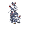 8x3wC 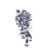 8x41C 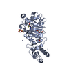 8x44C 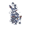 8x45C 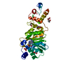 8x47C 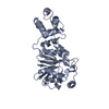 8x4gC 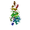 8x4iC 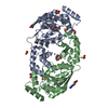 8x4lC  8x4oC 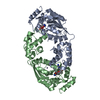 8x4pC C: citing same article ( |
|---|---|
| Similar structure data |
-Links
 PDBj
PDBj

- Assembly
| Deposited unit | 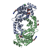
| ||||||||
|---|---|---|---|---|---|---|---|---|---|
| 1 |
| ||||||||
| Unit cell |
|
-Components
-Protein , 1 types, 2 molecules AB
| #1: Protein | Mass: 33365.117 Da / Num. of mol.: 2 Source method: isolated from a genetically manipulated source Source: (gene. exp.) Pyrococcus horikoshii OT3 (archaea) / Strain: OT3 / Gene: DIMT1 / Plasmid: pET28a / Production host:  References: UniProt: O59487, 16S rRNA (adenine1518-N6/adenine1519-N6)-dimethyltransferase |
|---|
-Non-polymers , 8 types, 251 molecules 


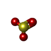


| #2: Chemical |  Mass: 381.387 Da / Num. of mol.: 2 / Source method: obtained synthetically / Formula: C15H23N7O5 / Feature type: SUBJECT OF INVESTIGATION Mass: 381.387 Da / Num. of mol.: 2 / Source method: obtained synthetically / Formula: C15H23N7O5 / Feature type: SUBJECT OF INVESTIGATION#3: Chemical | ChemComp-ZN /  Mass: 65.409 Da / Num. of mol.: 6 / Source method: obtained synthetically / Formula: Zn Mass: 65.409 Da / Num. of mol.: 6 / Source method: obtained synthetically / Formula: Zn#4: Chemical |  Mass: 35.453 Da / Num. of mol.: 2 / Source method: obtained synthetically / Formula: Cl Mass: 35.453 Da / Num. of mol.: 2 / Source method: obtained synthetically / Formula: Cl#5: Chemical |  Mass: 106.120 Da / Num. of mol.: 2 / Source method: obtained synthetically / Formula: C4H10O3 Mass: 106.120 Da / Num. of mol.: 2 / Source method: obtained synthetically / Formula: C4H10O3#6: Chemical |  Type: L-peptide linking / Mass: 175.209 Da / Num. of mol.: 2 / Source method: obtained synthetically / Formula: C6H15N4O2 Type: L-peptide linking / Mass: 175.209 Da / Num. of mol.: 2 / Source method: obtained synthetically / Formula: C6H15N4O2#7: Chemical | ChemComp-SO3 / |  Mass: 80.063 Da / Num. of mol.: 1 / Source method: obtained synthetically / Formula: SO3 Mass: 80.063 Da / Num. of mol.: 1 / Source method: obtained synthetically / Formula: SO3#8: Chemical | ChemComp-EDO / |  Mass: 62.068 Da / Num. of mol.: 1 / Source method: obtained synthetically / Formula: C2H6O2 Mass: 62.068 Da / Num. of mol.: 1 / Source method: obtained synthetically / Formula: C2H6O2#9: Water | ChemComp-HOH / | Mass: 18.015 Da / Num. of mol.: 235 / Source method: isolated from a natural source / Formula: H2O |
|---|
-Details
| Has ligand of interest | Y |
|---|---|
| Has protein modification | N |
-Experimental details
-Experiment
| Experiment | Method: X-RAY DIFFRACTION / Number of used crystals: 1 |
|---|
- Sample preparation
| Crystal | Density Matthews: 2.54 Å3/Da / Density % sol: 51.62 % / Description: C-centered Monoclinic |
|---|---|
| Crystal grow | Temperature: 293 K / Method: microbatch / pH: 6.5 Details: 0.2 M Zinc Acetate Dihydrate, 0.1 M Sodium Cacodylate Trihydrate pH 6.5, 18% (w/v) PEG 8000 |
-Data collection
| Diffraction | Mean temperature: 100 K / Serial crystal experiment: N |
|---|---|
| Diffraction source | Source: ROTATING ANODE / Type: RIGAKU MICROMAX-007 HF / Wavelength: 1.5418 Å |
| Detector | Type: RIGAKU RAXIS IV++ / Detector: IMAGE PLATE / Date: May 16, 2022 / Details: VariMax HF |
| Radiation | Monochromator: Nickel filter / Protocol: SINGLE WAVELENGTH / Monochromatic (M) / Laue (L): M / Scattering type: x-ray |
| Radiation wavelength | Wavelength: 1.5418 Å / Relative weight: 1 |
| Reflection | Resolution: 2.2→75.94 Å / Num. obs: 36508 / % possible obs: 100 % / Redundancy: 4.4 % / CC1/2: 0.994 / Rmerge(I) obs: 0.111 / Rpim(I) all: 0.059 / Rrim(I) all: 0.126 / Χ2: 0.58 / Net I/σ(I): 6.3 / Num. measured all: 161859 |
| Reflection shell | Resolution: 2.2→2.27 Å / % possible obs: 100 % / Redundancy: 4.3 % / Rmerge(I) obs: 0.554 / Num. measured all: 13702 / Num. unique obs: 3157 / CC1/2: 0.866 / Rpim(I) all: 0.302 / Rrim(I) all: 0.633 / Χ2: 0.57 / Net I/σ(I) obs: 2 |
- Processing
| Software |
| ||||||||||||||||||||||||||||||||||||||||||||||||||||||||||||||||||||||||||||||||||||||||||||||||||||||||||||||||||||||||||||||||||||||||||||||||||||||||||||||||||||||||||||||||||||||
|---|---|---|---|---|---|---|---|---|---|---|---|---|---|---|---|---|---|---|---|---|---|---|---|---|---|---|---|---|---|---|---|---|---|---|---|---|---|---|---|---|---|---|---|---|---|---|---|---|---|---|---|---|---|---|---|---|---|---|---|---|---|---|---|---|---|---|---|---|---|---|---|---|---|---|---|---|---|---|---|---|---|---|---|---|---|---|---|---|---|---|---|---|---|---|---|---|---|---|---|---|---|---|---|---|---|---|---|---|---|---|---|---|---|---|---|---|---|---|---|---|---|---|---|---|---|---|---|---|---|---|---|---|---|---|---|---|---|---|---|---|---|---|---|---|---|---|---|---|---|---|---|---|---|---|---|---|---|---|---|---|---|---|---|---|---|---|---|---|---|---|---|---|---|---|---|---|---|---|---|---|---|---|---|
| Refinement | Method to determine structure: MOLECULAR REPLACEMENT / Resolution: 2.2→64.45 Å / Cor.coef. Fo:Fc: 0.944 / Cor.coef. Fo:Fc free: 0.918 / SU B: 16.223 / SU ML: 0.207 / Cross valid method: THROUGHOUT / ESU R: 0.26 / ESU R Free: 0.215 / Stereochemistry target values: MAXIMUM LIKELIHOOD / Details: HYDROGENS HAVE BEEN ADDED IN THE RIDING POSITIONS
| ||||||||||||||||||||||||||||||||||||||||||||||||||||||||||||||||||||||||||||||||||||||||||||||||||||||||||||||||||||||||||||||||||||||||||||||||||||||||||||||||||||||||||||||||||||||
| Solvent computation | Ion probe radii: 0.8 Å / Shrinkage radii: 0.8 Å / VDW probe radii: 1.2 Å / Solvent model: MASK | ||||||||||||||||||||||||||||||||||||||||||||||||||||||||||||||||||||||||||||||||||||||||||||||||||||||||||||||||||||||||||||||||||||||||||||||||||||||||||||||||||||||||||||||||||||||
| Displacement parameters | Biso mean: 42.284 Å2
| ||||||||||||||||||||||||||||||||||||||||||||||||||||||||||||||||||||||||||||||||||||||||||||||||||||||||||||||||||||||||||||||||||||||||||||||||||||||||||||||||||||||||||||||||||||||
| Refinement step | Cycle: 1 / Resolution: 2.2→64.45 Å
| ||||||||||||||||||||||||||||||||||||||||||||||||||||||||||||||||||||||||||||||||||||||||||||||||||||||||||||||||||||||||||||||||||||||||||||||||||||||||||||||||||||||||||||||||||||||
| Refine LS restraints |
|