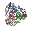
[English] 日本語
Yorodumi
- PDB-8wzs: Crystal structure of SRCRD11 of human DMBT1 from needle-shape crystal -

+
Open data


ID or keywords:

Loading...

-
Basic information

Entry
Database: PDB / ID: 8wzs
TitleCrystal structure of SRCRD11 of human DMBT1 from needle-shape crystal
ComponentsDeleted in malignant brain tumors 1 protein
KeywordsMETAL BINDING PROTEIN / Scavenger receptor cysteine-rich domain / Salivary scavenger and agglutinin
Function / homology
Function and homology information


induction of bacterial agglutination / zymogen granule membrane / Surfactant metabolism / zymogen binding / scavenger receptor activity / pattern recognition receptor activity / epithelial cell differentiation / extracellular matrix / defense response / phagocytic vesicle membrane ...induction of bacterial agglutination / zymogen granule membrane / Surfactant metabolism / zymogen binding / scavenger receptor activity / pattern recognition receptor activity / epithelial cell differentiation / extracellular matrix / defense response / phagocytic vesicle membrane / calcium-dependent protein binding / protein transport / defense response to Gram-negative bacterium / defense response to virus / defense response to Gram-positive bacterium / innate immune response / extracellular space / extracellular exosome / extracellular region / cytoplasm
Similarity search - Function
: / : / ZP-N domain / SRCR domain signature. / Zona pellucida domain, conserved site / ZP domain signature. / Zona pellucida, ZP-C domain / ZP-C domain / Zona pellucida (ZP) domain / Scavenger receptor cysteine-rich domain ...: / : / ZP-N domain / SRCR domain signature. / Zona pellucida domain, conserved site / ZP domain signature. / Zona pellucida, ZP-C domain / ZP-C domain / Zona pellucida (ZP) domain / Scavenger receptor cysteine-rich domain / ZP domain profile. / Zona pellucida domain / SRCR domain profile. / SRCR-like domain superfamily / Scavenger receptor Cys-rich / SRCR domain / CUB domain / Domain first found in C1r, C1s, uEGF, and bone morphogenetic protein. / CUB domain / CUB domain profile. / Spermadhesin, CUB domain superfamily
Similarity search - Domain/homology
Scavenger receptor cysteine-rich domain-containing protein DMBT1
Similarity search - Component
Biological speciesHomo sapiens (human)
MethodX-RAY DIFFRACTION / SYNCHROTRON / MOLECULAR REPLACEMENT / Resolution: 2.54 Å
AuthorsZhang, C. / Lu, P. / Nagata, K.
Funding support Japan, 1items
OrganizationGrant numberCountry
Japan Society for the Promotion of Science (JSPS)JP18H02151 Japan
CitationJournal: Protein J. / Year: 2024
Title: Refolding, Crystallization, and Crystal Structure Analysis of a Scavenger Receptor Cysteine-Rich Domain of Human Salivary Agglutinin Expressed in Escherichia coli.
Authors: Zhang, C. / Lu, P. / Wei, S. / Hu, C. / Miyoshi, M. / Okamoto, K. / Itoh, H. / Okuda, S. / Suzuki, M. / Kawakami, H. / Nagata, K.
History
DepositionNov 2, 2023Deposition site: PDBJ / Processing site: PDBJ
Revision 1.0Dec 11, 2024Provider: repository / Type: Initial release

-
Structure visualization

Structure viewerMolecule:
MolmilJmol/JSmol

Downloads & links

-
Assembly

Deposited unit
A: Deleted in malignant brain tumors 1 protein


Theoretical massNumber of molelcules
Total (without water)12,6801
Polymers12,6801
Non-polymers00
Water1,49583
1


  • Idetical with deposited unit
  • defined by author
  • Evidence: gel filtration
TypeNameSymmetry operationNumber
identity operation1_555x,y,z1
Unit cell
Length a, b, c (Å)71.114, 71.114, 55.387
Angle α, β, γ (deg.)90.00, 90.00, 120.00
Int Tables number154
Space group name H-MP3221

-
Components

#1: Protein Deleted in malignant brain tumors 1 protein / Glycoprotein 340 / Gp-340 / Hensin / Salivary agglutinin / SAG / Surfactant pulmonary-associated D- ...Glycoprotein 340 / Gp-340 / Hensin / Salivary agglutinin / SAG / Surfactant pulmonary-associated D-binding protein


Mass: 12679.798 Da / Num. of mol.: 1 / Fragment: SRCR domain 11
Source method: isolated from a genetically manipulated source
Source: (gene. exp.) Homo sapiens (human) / Gene: DMBT1, GP340 / Production host: Escherichia coli (E. coli) / References: UniProt: Q9UGM3
#2: Water ChemComp-HOH / water


Mass: 18.015 Da / Num. of mol.: 83 / Source method: isolated from a natural source / Formula: H2O
Has protein modificationY

-
Experimental details

-
Experiment

ExperimentMethod: X-RAY DIFFRACTION / Number of used crystals: 1

-
Sample preparation

CrystalDensity Matthews: 3.19 Å3/Da / Density % sol: 61.42 %
Crystal growTemperature: 293.15 K / Method: vapor diffusion, sitting drop / pH: 7
Details: 0.2 M Lithium citrate tribasic tetrahydrate, 25% w/v Polyethylene glycol 3,350

-
Data collection

DiffractionMean temperature: 100 K / Serial crystal experiment: N
Diffraction sourceSource: SYNCHROTRON / Site: Photon Factory / Beamline: AR-NE3A / Wavelength: 1 Å
DetectorType: DECTRIS PILATUS 2M-F / Detector: PIXEL / Date: Nov 24, 2021
RadiationProtocol: SINGLE WAVELENGTH / Monochromatic (M) / Laue (L): M / Scattering type: x-ray
Radiation wavelengthWavelength: 1 Å / Relative weight: 1
ReflectionResolution: 2.54→41.18 Å / Num. obs: 8237 / % possible obs: 99.73 % / Redundancy: 5.11 % / CC1/2: 0.998 / Net I/σ(I): 13.12
Reflection shellResolution: 2.54→7.51 Å / Num. unique obs: 1637 / CC1/2: 0.634

-
Processing

Software
NameVersionClassification
REFMAC5.8.0352refinement
XDSdata reduction
XDSdata scaling
MOLREPphasing
RefinementMethod to determine structure: MOLECULAR REPLACEMENT
Starting model: 6AS5
Resolution: 2.54→41.18 Å / Cor.coef. Fo:Fc: 0.964 / Cor.coef. Fo:Fc free: 0.93 / SU B: 9.105 / SU ML: 0.2 / Cross valid method: THROUGHOUT / ESU R: 0.324 / ESU R Free: 0.257 / Stereochemistry target values: MAXIMUM LIKELIHOOD / Details: HYDROGENS HAVE BEEN ADDED IN THE RIDING POSITIONS
RfactorNum. reflection% reflectionSelection details
Rfree0.24292 257 4.6 %RANDOM
Rwork0.17593 ---
obs0.17923 5307 99.73 %-
Solvent computationIon probe radii: 0.8 Å / Shrinkage radii: 0.8 Å / VDW probe radii: 1.2 Å / Solvent model: MASK
Displacement parametersBiso mean: 56.817 Å2
Baniso -1Baniso -2Baniso -3
1-0.03 Å20.02 Å2-0 Å2
2--0.03 Å20 Å2
3----0.1 Å2
Refinement stepCycle: 1 / Resolution: 2.54→41.18 Å
ProteinNucleic acidLigandSolventTotal
Num. atoms807 0 0 83 890
Refine LS restraints
Refine-IDTypeDev idealDev ideal targetNumber
X-RAY DIFFRACTIONr_bond_refined_d0.0080.011832
X-RAY DIFFRACTIONr_bond_other_d0.0010.016672
X-RAY DIFFRACTIONr_angle_refined_deg1.5851.6331135
X-RAY DIFFRACTIONr_angle_other_deg0.6161.5731566
X-RAY DIFFRACTIONr_dihedral_angle_1_deg9.1315106
X-RAY DIFFRACTIONr_dihedral_angle_2_deg7.75757
X-RAY DIFFRACTIONr_dihedral_angle_3_deg17.73510113
X-RAY DIFFRACTIONr_dihedral_angle_4_deg
X-RAY DIFFRACTIONr_chiral_restr0.0750.2114
X-RAY DIFFRACTIONr_gen_planes_refined0.0080.021003
X-RAY DIFFRACTIONr_gen_planes_other0.0010.02181
X-RAY DIFFRACTIONr_nbd_refined
X-RAY DIFFRACTIONr_nbd_other
X-RAY DIFFRACTIONr_nbtor_refined
X-RAY DIFFRACTIONr_nbtor_other
X-RAY DIFFRACTIONr_xyhbond_nbd_refined
X-RAY DIFFRACTIONr_xyhbond_nbd_other
X-RAY DIFFRACTIONr_metal_ion_refined
X-RAY DIFFRACTIONr_metal_ion_other
X-RAY DIFFRACTIONr_symmetry_vdw_refined
X-RAY DIFFRACTIONr_symmetry_vdw_other
X-RAY DIFFRACTIONr_symmetry_hbond_refined
X-RAY DIFFRACTIONr_symmetry_hbond_other
X-RAY DIFFRACTIONr_symmetry_metal_ion_refined
X-RAY DIFFRACTIONr_symmetry_metal_ion_other
X-RAY DIFFRACTIONr_mcbond_it5.8186.194427
X-RAY DIFFRACTIONr_mcbond_other5.8146.192427
X-RAY DIFFRACTIONr_mcangle_it7.7979.275532
X-RAY DIFFRACTIONr_mcangle_other7.7949.289533
X-RAY DIFFRACTIONr_scbond_it6.7746.764405
X-RAY DIFFRACTIONr_scbond_other6.6836.755404
X-RAY DIFFRACTIONr_scangle_it
X-RAY DIFFRACTIONr_scangle_other9.2949.956604
X-RAY DIFFRACTIONr_long_range_B_refined14.96994.2461037
X-RAY DIFFRACTIONr_long_range_B_other15.02792.0831018
X-RAY DIFFRACTIONr_rigid_bond_restr
X-RAY DIFFRACTIONr_sphericity_free
X-RAY DIFFRACTIONr_sphericity_bonded
LS refinement shellResolution: 2.54→2.606 Å / Total num. of bins used: 20
RfactorNum. reflection% reflection
Rfree0.463 24 -
Rwork0.231 358 -
obs--99.22 %

+
About Yorodumi

-
News

-
Feb 9, 2022. New format data for meta-information of EMDB entries

New format data for meta-information of EMDB entries

  • Version 3 of the EMDB header file is now the official format.
  • The previous official version 1.9 will be removed from the archive.

Related info.:EMDB header

External links:wwPDB to switch to version 3 of the EMDB data model

-
Aug 12, 2020. Covid-19 info

Covid-19 info

URL: https://pdbj.org/emnavi/covid19.php

New page: Covid-19 featured information page in EM Navigator.

Related info.:Covid-19 info / Mar 5, 2020. Novel coronavirus structure data

+
Mar 5, 2020. Novel coronavirus structure data

Novel coronavirus structure data

Related info.:Yorodumi Speices / Aug 12, 2020. Covid-19 info

External links:COVID-19 featured content - PDBj / Molecule of the Month (242):Coronavirus Proteases

+
Jan 31, 2019. EMDB accession codes are about to change! (news from PDBe EMDB page)

EMDB accession codes are about to change! (news from PDBe EMDB page)

  • The allocation of 4 digits for EMDB accession codes will soon come to an end. Whilst these codes will remain in use, new EMDB accession codes will include an additional digit and will expand incrementally as the available range of codes is exhausted. The current 4-digit format prefixed with “EMD-” (i.e. EMD-XXXX) will advance to a 5-digit format (i.e. EMD-XXXXX), and so on. It is currently estimated that the 4-digit codes will be depleted around Spring 2019, at which point the 5-digit format will come into force.
  • The EM Navigator/Yorodumi systems omit the EMD- prefix.

Related info.:Q: What is EMD? / ID/Accession-code notation in Yorodumi/EM Navigator

External links:EMDB Accession Codes are Changing Soon! / Contact to PDBj

+
Jul 12, 2017. Major update of PDB

Major update of PDB

  • wwPDB released updated PDB data conforming to the new PDBx/mmCIF dictionary.
  • This is a major update changing the version number from 4 to 5, and with Remediation, in which all the entries are updated.
  • In this update, many items about electron microscopy experimental information are reorganized (e.g. em_software).
  • Now, EM Navigator and Yorodumi are based on the updated data.

External links:wwPDB Remediation / Enriched Model Files Conforming to OneDep Data Standards Now Available in the PDB FTP Archive

-
Yorodumi

Thousand views of thousand structures

  • Yorodumi is a browser for structure data from EMDB, PDB, SASBDB, etc.
  • This page is also the successor to EM Navigator detail page, and also detail information page/front-end page for Omokage search.
  • The word "yorodu" (or yorozu) is an old Japanese word meaning "ten thousand". "mi" (miru) is to see.

Related info.:EMDB / PDB / SASBDB / Comparison of 3 databanks / Yorodumi Search / Aug 31, 2016. New EM Navigator & Yorodumi / Yorodumi Papers / Jmol/JSmol / Function and homology information / Changes in new EM Navigator and Yorodumi

Read more