 Movie
Movie Controller


+ Open data

- Basic information
| Entry | Database: PDB / ID: 8wzn | ||||||
|---|---|---|---|---|---|---|---|
| Title | ParkinK211N in complex with phospho NEDD8 | ||||||
 Components Components |
| ||||||
 Keywords Keywords | LIGASE / E3 ligase. | ||||||
| Function / homology |  Function and homology information Function and homology information: / positive regulation of retrograde transport, endosome to Golgi / regulation of lipid transport / positive regulation of neurotransmitter uptake / negative regulation of endoplasmic reticulum stress-induced neuron intrinsic apoptotic signaling pathway / negative regulation of spontaneous neurotransmitter secretion / negative regulation of intralumenal vesicle formation / regulation protein catabolic process at presynapse / cellular response to L-glutamine / : ...: / positive regulation of retrograde transport, endosome to Golgi / regulation of lipid transport / positive regulation of neurotransmitter uptake / negative regulation of endoplasmic reticulum stress-induced neuron intrinsic apoptotic signaling pathway / negative regulation of spontaneous neurotransmitter secretion / negative regulation of intralumenal vesicle formation / regulation protein catabolic process at presynapse / cellular response to L-glutamine / : / negative regulation of exosomal secretion / mitochondrion to lysosome vesicle-mediated transport / type 2 mitophagy / negative regulation of glucokinase activity / response to curcumin / negative regulation of mitochondrial fusion / cellular response to hydrogen sulfide / protein K29-linked ubiquitination / free ubiquitin chain polymerization / Lewy body / positive regulation of protein linear polyubiquitination / host-mediated suppression of viral genome replication / Parkin-FBXW7-Cul1 ubiquitin ligase complex / protein K27-linked ubiquitination / RBR-type E3 ubiquitin transferase / regulation of synaptic vesicle transport / positive regulation of mitochondrial fusion / negative regulation of actin filament bundle assembly / positive regulation of mitophagy / F-box domain binding / regulation of necroptotic process / mitochondrial fragmentation involved in apoptotic process / regulation of cellular response to oxidative stress / positive regulation of dendrite extension / negative regulation of excitatory postsynaptic potential / regulation of dopamine metabolic process / protein K6-linked ubiquitination / norepinephrine metabolic process / dopaminergic synapse / autophagy of mitochondrion / positive regulation of type 2 mitophagy / mitochondrion localization / protein localization to mitochondrion / positive regulation of tumor necrosis factor-mediated signaling pathway / cellular response to dopamine / positive regulation of proteasomal protein catabolic process / positive regulation of protein localization to membrane / mitochondrial fission / cellular response to toxic substance / negative regulation of intrinsic apoptotic signaling pathway by p53 class mediator / aggresome assembly / negative regulation of oxidative stress-induced neuron intrinsic apoptotic signaling pathway / cellular response to L-glutamate / regulation of mitochondrion organization / protein K11-linked ubiquitination / protein neddylation / ubiquitin conjugating enzyme binding / negative regulation of synaptic transmission, glutamatergic / regulation of canonical Wnt signaling pathway / positive regulation of mitochondrial membrane potential / negative regulation of JNK cascade / aggresome / regulation of reactive oxygen species metabolic process / response to corticosterone / positive regulation of mitochondrial fission / response to muscle activity / negative regulation of release of cytochrome c from mitochondria / ubiquitin-specific protease binding / startle response / dopamine metabolic process / dopamine uptake involved in synaptic transmission / regulation of dopamine secretion / positive regulation of ATP biosynthetic process / TGF-beta receptor signaling activates SMADs / negative regulation of reactive oxygen species metabolic process / regulation of glucose metabolic process / regulation of proteolysis / cullin family protein binding / regulation of protein ubiquitination / regulation of postsynapse assembly / protein deubiquitination / cellular response to unfolded protein / anatomical structure morphogenesis / protein K63-linked ubiquitination / regulation of synaptic vesicle endocytosis / protein monoubiquitination / ubiquitin ligase complex / negative regulation of mitochondrial fission / positive regulation of insulin secretion involved in cellular response to glucose stimulus / regulation of postsynaptic membrane neurotransmitter receptor levels / negative regulation of endoplasmic reticulum stress-induced intrinsic apoptotic signaling pathway / protein K48-linked ubiquitination / protein autoubiquitination / mitophagy / proteasomal protein catabolic process / phospholipase binding / negative regulation of reactive oxygen species biosynthetic process / heat shock protein binding / cellular response to manganese ion / ERAD pathway Similarity search - Function | ||||||
| Biological species |  Homo sapiens (human) Homo sapiens (human) | ||||||
| Method |  X-RAY DIFFRACTION / SYNCHROTRON / MOLECULAR REPLACEMENT / Resolution: 1.8 Å X-RAY DIFFRACTION / SYNCHROTRON / MOLECULAR REPLACEMENT / Resolution: 1.8 Å | ||||||
 Authors Authors | Lenka, D.R. / Kumar, A. | ||||||
| Funding support |  India, 1items India, 1items
| ||||||
 Citation Citation | Journal: Structure Title: Parkin in complex with phospho NEDD8 Authors: Lenka, D.R. / Kumar, A. | ||||||
| History |
|
- Structure visualization
| Structure viewer | Molecule: MolmilJmol/JSmol |
|---|
- Downloads & links
-Download
| PDBx/mmCIF format | 8wzn.cif.gz | 103.7 KB | Display | PDBx/mmCIF format |
|---|---|---|---|---|
| PDB format | pdb8wzn.ent.gz | 72.7 KB | Display | PDB format |
| PDBx/mmJSON format | 8wzn.json.gz | Tree view | PDBx/mmJSON format | |
| Others |  Other downloads Other downloads |
-Validation report
| Arichive directory | https://data.pdbj.org/pub/pdb/validation_reports/wz/8wznftp://data.pdbj.org/pub/pdb/validation_reports/wz/8wzn | HTTPS FTP |
|---|
-Related structure data
| Related structure data | 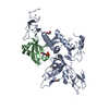 8wzoC 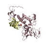 8ikvS S: Starting model for refinement C: citing same article ( |
|---|---|
| Similar structure data |
-Links
 PDBj
PDBj

- Assembly
| Deposited unit | 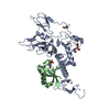
| ||||||||
|---|---|---|---|---|---|---|---|---|---|
| 1 |
| ||||||||
| Unit cell |
|
-Components
| #1: Protein | Mass: 36423.535 Da / Num. of mol.: 1 / Mutation: K211N Source method: isolated from a genetically manipulated source Source: (gene. exp.) Homo sapiens (human) / Gene: PRKN / Production host:  | ||||||||
|---|---|---|---|---|---|---|---|---|---|
| #2: Protein | Mass: 8741.034 Da / Num. of mol.: 1 Source method: isolated from a genetically manipulated source Source: (gene. exp.) Homo sapiens (human) / Gene: NEDD8 / Production host: | ||||||||
| #3: Chemical | ChemComp-ZN /   Mass: 65.409 Da / Num. of mol.: 8 / Source method: obtained synthetically / Formula: Zn Mass: 65.409 Da / Num. of mol.: 8 / Source method: obtained synthetically / Formula: Zn#4: Chemical |   Mass: 106.120 Da / Num. of mol.: 3 / Source method: obtained synthetically / Formula: C4H10O3 Mass: 106.120 Da / Num. of mol.: 3 / Source method: obtained synthetically / Formula: C4H10O3#5: Water | ChemComp-HOH / |  Mass: 18.015 Da / Num. of mol.: 319 / Source method: isolated from a natural source / Formula: H2O Mass: 18.015 Da / Num. of mol.: 319 / Source method: isolated from a natural source / Formula: H2OHas ligand of interest | N | Has protein modification | Y | |
-Experimental details
-Experiment
| Experiment | Method: X-RAY DIFFRACTION / Number of used crystals: 1 |
|---|
- Sample preparation
| Crystal | Density Matthews: 2 Å3/Da / Density % sol: 38.4 % |
|---|---|
| Crystal grow | Temperature: 291 K / Method: vapor diffusion / Details: 0.1 M, MMT, 7.0, 25% w/v, PEG 1500 |
-Data collection
| Diffraction | Mean temperature: 100 K / Serial crystal experiment: N |
|---|---|
| Diffraction source | Source: SYNCHROTRON / Site: ESRF  / Beamline: ID30B / Wavelength: 0.8731 Å / Beamline: ID30B / Wavelength: 0.8731 Å |
| Detector | Type: DECTRIS EIGER X 9M / Detector: PIXEL / Date: Sep 28, 2023 |
| Radiation | Protocol: SINGLE WAVELENGTH / Monochromatic (M) / Laue (L): M / Scattering type: x-ray |
| Radiation wavelength | Wavelength: 0.8731 Å / Relative weight: 1 |
| Reflection | Resolution: 1.8→39.86 Å / Num. obs: 32755 / % possible obs: 99.17 % / Redundancy: 4.5 % / CC1/2: 0.996 / Net I/σ(I): 10.25 |
| Reflection shell | Resolution: 1.8→1.864 Å / Num. unique obs: 3236 / CC1/2: 0.826 |
- Processing
| Software |
| |||||||||||||||||||||||||||||||||||||||||||||||||||||||||||||||||||||||||||||||||||||||||||||||||||||||||||||||||||||||||||||||||||||||||||||||||||||||||||||||||||||||||||||||||||||||||||||||||||||||||||||||||||||||||||||||||||||||
|---|---|---|---|---|---|---|---|---|---|---|---|---|---|---|---|---|---|---|---|---|---|---|---|---|---|---|---|---|---|---|---|---|---|---|---|---|---|---|---|---|---|---|---|---|---|---|---|---|---|---|---|---|---|---|---|---|---|---|---|---|---|---|---|---|---|---|---|---|---|---|---|---|---|---|---|---|---|---|---|---|---|---|---|---|---|---|---|---|---|---|---|---|---|---|---|---|---|---|---|---|---|---|---|---|---|---|---|---|---|---|---|---|---|---|---|---|---|---|---|---|---|---|---|---|---|---|---|---|---|---|---|---|---|---|---|---|---|---|---|---|---|---|---|---|---|---|---|---|---|---|---|---|---|---|---|---|---|---|---|---|---|---|---|---|---|---|---|---|---|---|---|---|---|---|---|---|---|---|---|---|---|---|---|---|---|---|---|---|---|---|---|---|---|---|---|---|---|---|---|---|---|---|---|---|---|---|---|---|---|---|---|---|---|---|---|---|---|---|---|---|---|---|---|---|---|---|---|---|---|---|---|---|
| Refinement | Method to determine structure: MOLECULAR REPLACEMENT Starting model: 8IKV Resolution: 1.8→39.86 Å / Cor.coef. Fo:Fc: 0.958 / Cor.coef. Fo:Fc free: 0.943 / WRfactor Rfree: 0.203 / WRfactor Rwork: 0.173 / SU B: 2.9 / SU ML: 0.089 / Average fsc free: 0.9321 / Average fsc work: 0.9415 / Cross valid method: FREE R-VALUE / ESU R: 0.144 / ESU R Free: 0.128 Details: Hydrogens have been added in their riding positions
| |||||||||||||||||||||||||||||||||||||||||||||||||||||||||||||||||||||||||||||||||||||||||||||||||||||||||||||||||||||||||||||||||||||||||||||||||||||||||||||||||||||||||||||||||||||||||||||||||||||||||||||||||||||||||||||||||||||||
| Solvent computation | Ion probe radii: 0.8 Å / Shrinkage radii: 0.8 Å / VDW probe radii: 1.2 Å / Solvent model: MASK BULK SOLVENT | |||||||||||||||||||||||||||||||||||||||||||||||||||||||||||||||||||||||||||||||||||||||||||||||||||||||||||||||||||||||||||||||||||||||||||||||||||||||||||||||||||||||||||||||||||||||||||||||||||||||||||||||||||||||||||||||||||||||
| Displacement parameters | Biso mean: 22.367 Å2
| |||||||||||||||||||||||||||||||||||||||||||||||||||||||||||||||||||||||||||||||||||||||||||||||||||||||||||||||||||||||||||||||||||||||||||||||||||||||||||||||||||||||||||||||||||||||||||||||||||||||||||||||||||||||||||||||||||||||
| Refinement step | Cycle: LAST / Resolution: 1.8→39.86 Å
| |||||||||||||||||||||||||||||||||||||||||||||||||||||||||||||||||||||||||||||||||||||||||||||||||||||||||||||||||||||||||||||||||||||||||||||||||||||||||||||||||||||||||||||||||||||||||||||||||||||||||||||||||||||||||||||||||||||||
| Refine LS restraints |
| |||||||||||||||||||||||||||||||||||||||||||||||||||||||||||||||||||||||||||||||||||||||||||||||||||||||||||||||||||||||||||||||||||||||||||||||||||||||||||||||||||||||||||||||||||||||||||||||||||||||||||||||||||||||||||||||||||||||
| LS refinement shell | Refine-ID: X-RAY DIFFRACTION / Total num. of bins used: 20
|
