 Movie
Movie Controller


+ Open data

- Basic information
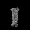
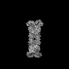
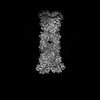
| Entry | Database: PDB / ID: 8wyc | ||||||
|---|---|---|---|---|---|---|---|
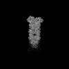
| Title | Cryo-EM structure of DSR2 (H171A)-tube-NAD+ (partial) complex | ||||||
 Components Components |
| ||||||
 Keywords Keywords | ANTIVIRAL PROTEIN / Phage defense proteins | ||||||
| Function / homology | : / YomU/Tube protein family / SIR2-like domain / SIR2-like domain / DHS-like NAD/FAD-binding domain superfamily / NICOTINAMIDE-ADENINE-DINUCLEOTIDE / Uncharacterized protein / SIR2-like domain-containing protein Function and homology information Function and homology information | ||||||
| Biological species |   Bacillus phage SPR (virus) Bacillus phage SPR (virus) | ||||||
| Method | ELECTRON MICROSCOPY / single particle reconstruction / cryo EM / Resolution: 3 Å | ||||||
 Authors Authors | Zhang, J.T. / Jia, N. / Liu, X.Y. | ||||||
| Funding support | 1items
| ||||||
 Citation Citation | Journal: Nat Commun / Year: 2024 Title: Structural basis for phage-mediated activation and repression of bacterial DSR2 anti-phage defense system. Authors: Jun-Tao Zhang / Xiao-Yu Liu / Zhuolin Li / Xin-Yang Wei / Xin-Yi Song / Ning Cui / Jirui Zhong / Hongchun Li / Ning Jia /  Abstract: Silent information regulator 2 (Sir2) proteins typically catalyze NAD-dependent protein deacetylation. The recently identified bacterial Sir2 domain-containing protein, defense-associated sirtuin 2 ...Silent information regulator 2 (Sir2) proteins typically catalyze NAD-dependent protein deacetylation. The recently identified bacterial Sir2 domain-containing protein, defense-associated sirtuin 2 (DSR2), recognizes the phage tail tube and depletes NAD to abort phage propagation, which is counteracted by the phage-encoded DSR anti-defense 1 (DSAD1), but their molecular mechanisms remain unclear. Here, we determine cryo-EM structures of inactive DSR2 in its apo form, DSR2-DSAD1 and DSR2-DSAD1-NAD, as well as active DSR2-tube and DSR2-tube-NAD complexes. DSR2 forms a tetramer with its C-terminal sensor domains (CTDs) in two distinct conformations: CTD or CTD. Monomeric, rather than oligomeric, tail tube proteins preferentially bind to CTD and activate Sir2 for NAD hydrolysis. DSAD1 binding to CTD allosterically inhibits tube binding and tube-mediated DSR2 activation. Our findings provide mechanistic insight into DSR2 assembly, tube-mediated DSR2 activation, and DSAD1-mediated inhibition and NAD substrate catalysis in bacterial DSR2 anti-phage defense systems. | ||||||
| History |
|
- Structure visualization
| Structure viewer | Molecule: MolmilJmol/JSmol |
|---|
- Downloads & links
-Download
| PDBx/mmCIF format | 8wyc.cif.gz | 574.4 KB | Display | PDBx/mmCIF format |
|---|---|---|---|---|
| PDB format | pdb8wyc.ent.gz | 454.6 KB | Display | PDB format |
| PDBx/mmJSON format | 8wyc.json.gz | Tree view | PDBx/mmJSON format | |
| Others |  Other downloads Other downloads |
-Validation report
| Arichive directory | https://data.pdbj.org/pub/pdb/validation_reports/wy/8wycftp://data.pdbj.org/pub/pdb/validation_reports/wy/8wyc | HTTPS FTP |
|---|
-Related structure data
| Related structure data | 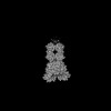 37923MC 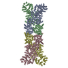 8wy8C 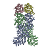 8wy9C 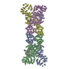 8wyaC 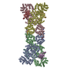 8wybC 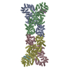 8wydC 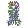 8wyeC 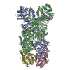 8wyfC M: map data used to model this data C: citing same article ( |
|---|---|
| Similar structure data |

-Links
 PDBj
PDBj

- Assembly
| Deposited unit | 
|
|---|---|
| 1 |
|
-Components
| #1: Protein | Mass: 118568.727 Da / Num. of mol.: 4 / Mutation: H171A Source method: isolated from a genetically manipulated source Source: (gene. exp.) #2: Protein | Mass: 29304.701 Da / Num. of mol.: 2 Source method: isolated from a genetically manipulated source Source: (gene. exp.) Bacillus phage SPR (virus) / Gene: B4122_1986 / Production host: #3: Chemical | ChemComp-NAD /   Mass: 663.425 Da / Num. of mol.: 4 / Source method: obtained synthetically / Formula: C21H27N7O14P2 / Feature type: SUBJECT OF INVESTIGATION / Comment: NAD*YM Mass: 663.425 Da / Num. of mol.: 4 / Source method: obtained synthetically / Formula: C21H27N7O14P2 / Feature type: SUBJECT OF INVESTIGATION / Comment: NAD*YMHas ligand of interest | Y | |
|---|
-Experimental details
-Experiment
| Experiment | Method: ELECTRON MICROSCOPY |
|---|---|
| EM experiment | Aggregation state: PARTICLE / 3D reconstruction method: single particle reconstruction |
- Sample preparation
| Component | Name: CryoEM structure of DSR2 (H171A)-Tube NAD+ (half) complex Type: COMPLEX / Entity ID: #1-#2 / Source: RECOMBINANT |
|---|---|
| Source (natural) | Organism: |
| Source (recombinant) | Organism: |
| Buffer solution | pH: 7.5 |
| Specimen | Embedding applied: NO / Shadowing applied: NO / Staining applied: NO / Vitrification applied: YES |
| Vitrification | Cryogen name: ETHANE |
- Electron microscopy imaging
| Experimental equipment |  Model: Titan Krios / Image courtesy: FEI Company |
|---|---|
| Microscopy | Model: FEI TITAN KRIOS |
| Electron gun | Electron source:  FIELD EMISSION GUN / Accelerating voltage: 300 kV / Illumination mode: SPOT SCAN FIELD EMISSION GUN / Accelerating voltage: 300 kV / Illumination mode: SPOT SCAN |
| Electron lens | Mode: BRIGHT FIELD / Nominal defocus max: 2500 nm / Nominal defocus min: 1500 nm / Cs: 2.7 mm / C2 aperture diameter: 50 µm |
| Image recording | Electron dose: 50 e/Å2 / Film or detector model: GATAN K3 (6k x 4k) |
- Processing
| CTF correction | Type: PHASE FLIPPING ONLY |
|---|---|
| 3D reconstruction | Resolution: 3 Å / Resolution method: FSC 0.143 CUT-OFF / Num. of particles: 85495 / Symmetry type: POINT |
