 Movie
Movie Controller

[English] 日本語
 Yorodumi
- PDB-8wsv: Pre-binding structure of HosA transcriptional regulator from ente... -
+ Open data

- Basic information
| Entry | Database: PDB / ID: 8wsv | ||||||
|---|---|---|---|---|---|---|---|
| Title | Pre-binding structure of HosA transcriptional regulator from enteropathogenic Escherichia coli O127:H6 (strain E2348/69) in presence of sub optimal concentration of 4-hydroxy benzoic acid | ||||||
 Components Components | Transcriptional regulator HosA | ||||||
 Keywords Keywords | DNA BINDING PROTEIN / Antibiotic resistance / MarR transcription factor / HosA / Enteropathogenic Escherichia coli | ||||||
| Function / homology |  Function and homology information Function and homology information | ||||||
| Biological species |  | ||||||
| Method |  X-RAY DIFFRACTION / MOLECULAR REPLACEMENT / Resolution: 2.01 Å X-RAY DIFFRACTION / MOLECULAR REPLACEMENT / Resolution: 2.01 Å | ||||||
 Authors Authors | Goswami, A. / Raju, R. / Kasarla, M. / Ullah, S. | ||||||
| Funding support | 1items
| ||||||
 Citation Citation | Journal: Biorxiv / Year: 2024 Title: Pre-binding structure of HosA transcriptional regulator from enteropathogenic Escherichia coli O127:H6 (strain E2348/69) in presence of sub optimal concentration of 4-hydroxy benzoic acid Authors: Arpita, G. / Rukmini, R. / Mallesham, K. / Samee, U. | ||||||
| History |
|
- Structure visualization
| Structure viewer | Molecule: MolmilJmol/JSmol |
|---|
- Downloads & links
-Download
| PDBx/mmCIF format | 8wsv.cif.gz | 45 KB | Display | PDBx/mmCIF format |
|---|---|---|---|---|
| PDB format | pdb8wsv.ent.gz | 29.1 KB | Display | PDB format |
| PDBx/mmJSON format | 8wsv.json.gz | Tree view | PDBx/mmJSON format | |
| Others |  Other downloads Other downloads |
-Validation report
| Arichive directory | https://data.pdbj.org/pub/pdb/validation_reports/ws/8wsvftp://data.pdbj.org/pub/pdb/validation_reports/ws/8wsv | HTTPS FTP |
|---|
-Related structure data
| Related structure data |  8xb7C  8pq4S  8kg0 S: Starting model for refinement C: citing same article ( |
|---|---|
| Similar structure data |

-Links
 PDBj
PDBj- Assembly
| Deposited unit | 
| ||||||||
|---|---|---|---|---|---|---|---|---|---|
| 1 | 
| ||||||||
| Unit cell |
| ||||||||
| Components on special symmetry positions |
|
-Components
| #1: Protein | Mass: 16592.092 Da / Num. of mol.: 1 Source method: isolated from a genetically manipulated source Details: HosA protein Source: (gene. exp.) Gene: hosA / Production host: |
|---|---|
| #2: Chemical | ChemComp-PHB / 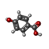  Mass: 138.121 Da / Num. of mol.: 1 / Source method: isolated from a natural source / Formula: C7H6O3 / Feature type: SUBJECT OF INVESTIGATION Mass: 138.121 Da / Num. of mol.: 1 / Source method: isolated from a natural source / Formula: C7H6O3 / Feature type: SUBJECT OF INVESTIGATION |
| #3: Water | ChemComp-HOH /  Mass: 18.015 Da / Num. of mol.: 138 / Source method: isolated from a natural source / Formula: H2O Mass: 18.015 Da / Num. of mol.: 138 / Source method: isolated from a natural source / Formula: H2O |
| Has ligand of interest | Y |
-Experimental details
-Experiment
| Experiment | Method: X-RAY DIFFRACTION / Number of used crystals: 1 |
|---|
- Sample preparation
| Crystal | Density Matthews: 3.52 Å3/Da / Density % sol: 65.06 % |
|---|---|
| Crystal grow | Temperature: 277.15 K / Method: microbatch / pH: 6.2 Details: Sodium phosphate dibasic/ Potassium phosphate monobasic, Sodium chloride, PEG 200 |
-Data collection
| Diffraction | Mean temperature: 100 K / Serial crystal experiment: N |
|---|---|
| Diffraction source | Source: ROTATING ANODE / Type: RIGAKU FR-E+ SUPERBRIGHT / Wavelength: 1.54187 Å |
| Detector | Type: RIGAKU RAXIS IV++ / Detector: IMAGE PLATE / Date: Aug 8, 2023 / Details: Tube |
| Radiation | Protocol: SINGLE WAVELENGTH / Monochromatic (M) / Laue (L): M / Scattering type: x-ray |
| Radiation wavelength | Wavelength: 1.54187 Å / Relative weight: 1 |
| Reflection | Resolution: 2.01→67.47 Å / Num. obs: 14952 / % possible obs: 98.1 % / Redundancy: 7 % / Biso Wilson estimate: 13.28 Å2 / CC1/2: 0.997 / Rmerge(I) obs: 0.065 / Rpim(I) all: 0.026 / Rrim(I) all: 0.07 / Net I/σ(I): 21 |
| Reflection shell | Resolution: 2.01→2.12 Å / Redundancy: 7 % / Rmerge(I) obs: 0.116 / Mean I/σ(I) obs: 13 / Num. unique obs: 2096 / CC1/2: 0.992 / Rpim(I) all: 0.046 / Rrim(I) all: 0.125 / % possible all: 96.6 |
- Processing
| Software |
| ||||||||||||||||||||||||||||||||||||||||||||||||||||||||||||||||||||||||||||||||||||
|---|---|---|---|---|---|---|---|---|---|---|---|---|---|---|---|---|---|---|---|---|---|---|---|---|---|---|---|---|---|---|---|---|---|---|---|---|---|---|---|---|---|---|---|---|---|---|---|---|---|---|---|---|---|---|---|---|---|---|---|---|---|---|---|---|---|---|---|---|---|---|---|---|---|---|---|---|---|---|---|---|---|---|---|---|---|
| Refinement | Method to determine structure: MOLECULAR REPLACEMENT Starting model: 8PQ4 Resolution: 2.01→42.65 Å / SU ML: 0.21 / Cross valid method: FREE R-VALUE / σ(F): 1.34 / Phase error: 23.85 / Stereochemistry target values: ML Details: TLS refinement along with occupancy was carried out for few steps. Once the partial density of the ligand was clear which aligned perfectly with ligand present in 8AGA, PHB was modeled with ...Details: TLS refinement along with occupancy was carried out for few steps. Once the partial density of the ligand was clear which aligned perfectly with ligand present in 8AGA, PHB was modeled with partial occupancy and the structure was refined using 8AGA as reference model and TLS = off.
| ||||||||||||||||||||||||||||||||||||||||||||||||||||||||||||||||||||||||||||||||||||
| Solvent computation | Shrinkage radii: 0.9 Å / VDW probe radii: 1.11 Å / Solvent model: FLAT BULK SOLVENT MODEL | ||||||||||||||||||||||||||||||||||||||||||||||||||||||||||||||||||||||||||||||||||||
| Displacement parameters | Biso mean: 16.45 Å2 | ||||||||||||||||||||||||||||||||||||||||||||||||||||||||||||||||||||||||||||||||||||
| Refinement step | Cycle: LAST / Resolution: 2.01→42.65 Å
| ||||||||||||||||||||||||||||||||||||||||||||||||||||||||||||||||||||||||||||||||||||
| Refine LS restraints |
| ||||||||||||||||||||||||||||||||||||||||||||||||||||||||||||||||||||||||||||||||||||
| LS refinement shell |
|
