 Movie
Movie Controller

[English] 日本語
 Yorodumi
- PDB-8wpz: Cryo-ET structure of RuBisCO at 3.9 angstroms from Synechococcus ... -
+ Open data

- Basic information
| Entry | Database: PDB / ID: 8wpz | |||||||||||||||||||||||||||||||||||||||||||||||||||||||||
|---|---|---|---|---|---|---|---|---|---|---|---|---|---|---|---|---|---|---|---|---|---|---|---|---|---|---|---|---|---|---|---|---|---|---|---|---|---|---|---|---|---|---|---|---|---|---|---|---|---|---|---|---|---|---|---|---|---|---|
| Title | Cryo-ET structure of RuBisCO at 3.9 angstroms from Synechococcus elongatus PCC 7942 | |||||||||||||||||||||||||||||||||||||||||||||||||||||||||
 Components Components |
| |||||||||||||||||||||||||||||||||||||||||||||||||||||||||
 Keywords Keywords | PHOTOSYNTHESIS / carboxysome / RuBisCO / cryo-et | |||||||||||||||||||||||||||||||||||||||||||||||||||||||||
| Function / homology |  Function and homology information Function and homology informationphotorespiration / carboxysome / ribulose-bisphosphate carboxylase / ribulose-bisphosphate carboxylase activity / reductive pentose-phosphate cycle / monooxygenase activity / magnesium ion binding Similarity search - Function | |||||||||||||||||||||||||||||||||||||||||||||||||||||||||
| Biological species |  Synechococcus elongatus PCC 7942 = FACHB-805 (bacteria) Synechococcus elongatus PCC 7942 = FACHB-805 (bacteria) | |||||||||||||||||||||||||||||||||||||||||||||||||||||||||
| Method | ELECTRON MICROSCOPY / subtomogram averaging / cryo EM / Resolution: 3.9 Å | |||||||||||||||||||||||||||||||||||||||||||||||||||||||||
 Authors Authors | Kong, W.W. / Jiang, Y.L. / Zhou, C.Z. | |||||||||||||||||||||||||||||||||||||||||||||||||||||||||
| Funding support |  China, 1items China, 1items
| |||||||||||||||||||||||||||||||||||||||||||||||||||||||||
 Citation Citation | Journal: Structure / Year: 2024 Title: Cryo-electron tomography reveals the packaging pattern of RuBisCOs in Synechococcus β-carboxysome. Authors: Wen-Wen Kong / Yun Zhu / Heng-Rui Zhao / Kang Du / Rui-Qian Zhou / Bo Li / Feng Yang / Pu Hou / Xia-He Huang / Yuxing Chen / Ying-Chun Wang / Fei Sun / Yong-Liang Jiang / Cong-Zhao Zhou / Abstract: Carboxysomes are large self-assembled microcompartments that serve as the central machinery of a CO-concentrating mechanism (CCM). Biogenesis of carboxysome requires the fine organization of ...Carboxysomes are large self-assembled microcompartments that serve as the central machinery of a CO-concentrating mechanism (CCM). Biogenesis of carboxysome requires the fine organization of thousands of individual proteins; however, the packaging pattern of internal RuBisCOs remains largely unknown. Here we purified the intact β-carboxysomes from Synechococcus elongatus PCC 7942 and identified the protein components by mass spectrometry. Cryo-electron tomography combined with subtomogram averaging revealed the general organization pattern of internal RuBisCOs, in which the adjacent RuBisCOs are mainly arranged in three distinct manners: head-to-head, head-to-side, and side-by-side. The RuBisCOs in the outermost layer are regularly aligned along the shell, the majority of which directly interact with the shell. Moreover, statistical analysis enabled us to propose an ideal packaging model of RuBisCOs in the β-carboxysome. These results provide new insights into the biogenesis of β-carboxysomes and also advance our understanding of the efficient carbon fixation functionality of carboxysomes. | |||||||||||||||||||||||||||||||||||||||||||||||||||||||||
| History |
|
- Structure visualization
| Structure viewer | Molecule: MolmilJmol/JSmol |
|---|
- Downloads & links
-Download
| PDBx/mmCIF format | 8wpz.cif.gz | 757.5 KB | Display | PDBx/mmCIF format |
|---|---|---|---|---|
| PDB format | pdb8wpz.ent.gz | 635.4 KB | Display | PDB format |
| PDBx/mmJSON format | 8wpz.json.gz | Tree view | PDBx/mmJSON format | |
| Others |  Other downloads Other downloads |
-Validation report
| Summary document | 8wpz_validation.pdf.gz | 459.5 KB | Display | wwPDB validaton report |
|---|---|---|---|---|
| Full document | 8wpz_full_validation.pdf.gz | 535.1 KB | Display | |
| Data in XML | 8wpz_validation.xml.gz | 89.9 KB | Display | |
| Data in CIF | 8wpz_validation.cif.gz | 136.1 KB | Display | |
| Arichive directory | https://data.pdbj.org/pub/pdb/validation_reports/wp/8wpzftp://data.pdbj.org/pub/pdb/validation_reports/wp/8wpz | HTTPS FTP |
-Related structure data
| Related structure data |  37727MC M: map data used to model this data C: citing same article ( |
|---|---|
| Similar structure data |


-Links
 PDBj
PDBj
- Assembly
| Deposited unit | 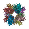
|
|---|---|
| 1 |
|
-Components
| #1: Protein | Mass: 13349.196 Da / Num. of mol.: 8 / Source method: isolated from a natural source Source: (natural) Synechococcus elongatus PCC 7942 = FACHB-805 (bacteria)References: UniProt: Q31NB2 #2: Protein | Mass: 52516.605 Da / Num. of mol.: 8 / Source method: isolated from a natural source Source: (natural) Synechococcus elongatus PCC 7942 = FACHB-805 (bacteria)References: UniProt: Q31NB3, ribulose-bisphosphate carboxylase Has protein modification | Y | |
|---|
-Experimental details
-Experiment
| Experiment | Method: ELECTRON MICROSCOPY |
|---|---|
| EM experiment | Aggregation state: PARTICLE / 3D reconstruction method: subtomogram averaging |
- Sample preparation
| Component | Name: RuBisCO / Type: COMPLEX / Entity ID: all / Source: NATURAL |
|---|---|
| Source (natural) | Organism: Synechococcus elongatus (strain ATCC 33912 / PCC 7942 / FACHB-805) (bacteria) |
| Details of virus | Empty: NO / Enveloped: NO / Isolate: OTHER / Type: VIRION |
| Natural host | Organism: Nostoc sp. PCC 7120 = FACHB-418 |
| Buffer solution | pH: 8 |
| Specimen | Embedding applied: NO / Shadowing applied: NO / Staining applied: NO / Vitrification applied: YES |
| Vitrification | Cryogen name: ETHANE / Humidity: 100 % / Chamber temperature: 300 K |
- Electron microscopy imaging
| Experimental equipment |  Model: Titan Krios / Image courtesy: FEI Company |
|---|---|
| Microscopy | Model: FEI TITAN KRIOS |
| Electron gun | Electron source:  FIELD EMISSION GUN / Accelerating voltage: 300 kV / Illumination mode: SPOT SCAN FIELD EMISSION GUN / Accelerating voltage: 300 kV / Illumination mode: SPOT SCAN |
| Electron lens | Mode: BRIGHT FIELD / Nominal defocus max: 3000 nm / Nominal defocus min: 1000 nm |
| Image recording | Electron dose: 3.5 e/Å2 / Avg electron dose per subtomogram: 145 e/Å2 / Film or detector model: GATAN K2 SUMMIT (4k x 4k) |
- Processing
| EM software |
| ||||||||||||||||||||||||
|---|---|---|---|---|---|---|---|---|---|---|---|---|---|---|---|---|---|---|---|---|---|---|---|---|---|
| CTF correction | Type: PHASE FLIPPING ONLY | ||||||||||||||||||||||||
| 3D reconstruction | Resolution: 3.9 Å / Resolution method: FSC 0.143 CUT-OFF / Num. of particles: 2700 / Symmetry type: POINT | ||||||||||||||||||||||||
| EM volume selection | Num. of tomograms: 88 / Num. of volumes extracted: 29000 | ||||||||||||||||||||||||
| Refine LS restraints |
|
