[English] 日本語
Yorodumi
- PDB-8wnf: Crystal structure of H. pylori isoleucyl-tRNA synthetase (HpIleRS... -

+
Open data


ID or keywords:

Loading...

-
Basic information

Entry
Database: PDB / ID: 8wnf
TitleCrystal structure of H. pylori isoleucyl-tRNA synthetase (HpIleRS) in apo form
ComponentsIsoleucine--tRNA ligase
KeywordsLIGASE / isoleucyl-tRNA synthetase / Helicobacter pylori / HpIleRS / IleRS / aminoacylation
Function / homology
Function and homology information


isoleucine-tRNA ligase / isoleucine-tRNA ligase activity / isoleucyl-tRNA aminoacylation / aminoacyl-tRNA editing activity / tRNA binding / zinc ion binding / ATP binding / cytoplasm
Similarity search - Function
Isoleucine-tRNA ligase, type 1 / Isoleucyl tRNA synthetase type 1, anticodon-binding domain / Isoleucine-tRNA ligase / Aminoacyl-tRNA synthetase, class Ia / tRNA synthetases class I (I, L, M and V) / Valyl/Leucyl/Isoleucyl-tRNA synthetase, editing domain / Methionyl/Valyl/Leucyl/Isoleucyl-tRNA synthetase, anticodon-binding / Anticodon-binding domain of tRNA ligase / Aminoacyl-tRNA synthetase, class Ia, anticodon-binding / Aminoacyl-tRNA synthetase, class I, conserved site ...Isoleucine-tRNA ligase, type 1 / Isoleucyl tRNA synthetase type 1, anticodon-binding domain / Isoleucine-tRNA ligase / Aminoacyl-tRNA synthetase, class Ia / tRNA synthetases class I (I, L, M and V) / Valyl/Leucyl/Isoleucyl-tRNA synthetase, editing domain / Methionyl/Valyl/Leucyl/Isoleucyl-tRNA synthetase, anticodon-binding / Anticodon-binding domain of tRNA ligase / Aminoacyl-tRNA synthetase, class Ia, anticodon-binding / Aminoacyl-tRNA synthetase, class I, conserved site / Aminoacyl-transfer RNA synthetases class-I signature. / Rossmann-like alpha/beta/alpha sandwich fold
Similarity search - Domain/homology
ACETATE ION / Isoleucine--tRNA ligase
Similarity search - Component
Biological speciesHelicobacter pylori (bacteria)
MethodX-RAY DIFFRACTION / SYNCHROTRON / SAD / Resolution: 1.9 Å
AuthorsGuo, Y. / Li, S. / Zhang, T.
Funding support China, 1items
OrganizationGrant numberCountry
National Natural Science Foundation of China (NSFC) China
CitationJournal: Febs Lett. / Year: 2024
Title: Structural basis for substrate and antibiotic recognition by Helicobacter pylori isoleucyl-tRNA synthetase.
Authors: Chen, X. / Guo, Y. / Shi, J. / Wang, Y. / Guo, X. / Wu, G. / Li, S. / Zhang, T.
History
DepositionOct 5, 2023Deposition site: PDBJ / Processing site: PDBC
Revision 1.0Feb 14, 2024Provider: repository / Type: Initial release
Revision 1.1Mar 27, 2024Group: Database references / Category: citation / citation_author
Item: _citation.journal_volume / _citation.page_first ..._citation.journal_volume / _citation.page_first / _citation.page_last / _citation_author.identifier_ORCID

-
Structure visualization

Structure viewerMolecule:
MolmilJmol/JSmol

Downloads & links

-
Assembly

Deposited unit
A: Isoleucine--tRNA ligase
hetero molecules


Theoretical massNumber of molelcules
Total (without water)107,5228
Polymers106,9371
Non-polymers5857
Water19,3481074
1


  • Idetical with deposited unit
  • defined by author&software
  • Evidence: gel filtration
TypeNameSymmetry operationNumber
identity operation1_555x,y,z1
Buried area230 Å2
ΔGint-0 kcal/mol
Surface area40430 Å2
MethodPISA
Unit cell
Length a, b, c (Å)52.623, 52.776, 103.254
Angle α, β, γ (deg.)92.58, 97.77, 103.71
Int Tables number1
Space group name H-MP1

-
Components

#1: Protein Isoleucine--tRNA ligase / Isoleucyl-tRNA synthetase / IleRS


Mass: 106936.977 Da / Num. of mol.: 1
Source method: isolated from a genetically manipulated source
Source: (gene. exp.) Helicobacter pylori (bacteria) / Gene: ileS, C2840_07400 / Production host: Escherichia coli (E. coli) / References: UniProt: A0A2J9KLI1, isoleucine-tRNA ligase
#2: Chemical
ChemComp-GOL / GLYCEROL / GLYCERIN / PROPANE-1,2,3-TRIOL


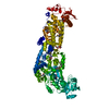
Mass: 92.094 Da / Num. of mol.: 5 / Source method: obtained synthetically / Formula: C3H8O3
#3: Chemical ChemComp-ZN / ZINC ION


Mass: 65.409 Da / Num. of mol.: 1 / Source method: obtained synthetically / Formula: Zn
#4: Chemical ChemComp-ACT / ACETATE ION


Mass: 59.044 Da / Num. of mol.: 1 / Source method: obtained synthetically / Formula: C2H3O2
#5: Water ChemComp-HOH / water


Mass: 18.015 Da / Num. of mol.: 1074 / Source method: isolated from a natural source / Formula: H2O
Has ligand of interestN

-
Experimental details

-
Experiment

ExperimentMethod: X-RAY DIFFRACTION / Number of used crystals: 1

-
Sample preparation

CrystalDensity Matthews: 2.59 Å3/Da / Density % sol: 52.51 %
Crystal growTemperature: 289 K / Method: vapor diffusion, hanging drop / pH: 7.5
Details: 0.1 M Hepes, pH 7.5, 2% (v/v) Tacsimate, and 20% (w/v) polyethylene glycol (PEG) 3,350

-
Data collection

DiffractionMean temperature: 100 K / Serial crystal experiment: N
Diffraction sourceSource: SYNCHROTRON / Site: SSRF / Beamline: BL19U1 / Wavelength: 0.9785 Å
DetectorType: DECTRIS PILATUS 6M / Detector: PIXEL / Date: Nov 7, 2019
RadiationProtocol: SINGLE WAVELENGTH / Monochromatic (M) / Laue (L): M / Scattering type: x-ray
Radiation wavelengthWavelength: 0.9785 Å / Relative weight: 1
ReflectionResolution: 1.9→48.31 Å / Num. obs: 81231 / % possible obs: 97.09 % / Redundancy: 3.4 % / CC1/2: 0.997 / Rmerge(I) obs: 0.0727 / Net I/σ(I): 12.3
Reflection shellResolution: 1.9→1.97 Å / Rmerge(I) obs: 1.53 / Num. unique obs: 7613 / CC1/2: 0.663 / % possible all: 90.2

-
Processing

Software
NameVersionClassification
REFMAC5.8.0238refinement
XDSdata reduction
XDSdata scaling
PHENIXphasing
RefinementMethod to determine structure: SAD / Resolution: 1.9→48.31 Å / Cor.coef. Fo:Fc: 0.966 / Cor.coef. Fo:Fc free: 0.947 / SU B: 2.669 / SU ML: 0.079 / Cross valid method: THROUGHOUT / ESU R: 0.131 / ESU R Free: 0.125 / Stereochemistry target values: MAXIMUM LIKELIHOOD
RfactorNum. reflection% reflectionSelection details
Rfree0.19096 4124 5.1 %RANDOM
Rwork0.1508 ---
obs0.15091 77107 97.08 %-
Solvent computationIon probe radii: 0.8 Å / Shrinkage radii: 0.8 Å / VDW probe radii: 1.2 Å / Solvent model: MASK
Displacement parametersBiso mean: 22.946 Å2
Baniso -1Baniso -2Baniso -3
1-0.21 Å2-0.01 Å2-0.09 Å2
2--0.09 Å2-0.19 Å2
3----0.22 Å2
Refinement stepCycle: 1 / Resolution: 1.9→48.31 Å
ProteinNucleic acidLigandSolventTotal
Num. atoms7464 0 35 1074 8573
Refine LS restraints
Refine-IDTypeDev idealDev ideal targetNumber
X-RAY DIFFRACTIONr_bond_refined_d0.010.0127661
X-RAY DIFFRACTIONr_bond_other_d
X-RAY DIFFRACTIONr_angle_refined_deg1.5791.63610331
X-RAY DIFFRACTIONr_angle_other_deg
X-RAY DIFFRACTIONr_dihedral_angle_1_deg5.7735917
X-RAY DIFFRACTIONr_dihedral_angle_2_deg33.8723.625400
X-RAY DIFFRACTIONr_dihedral_angle_3_deg14.1151374
X-RAY DIFFRACTIONr_dihedral_angle_4_deg16.241532
X-RAY DIFFRACTIONr_chiral_restr0.1080.2955
X-RAY DIFFRACTIONr_gen_planes_refined0.010.025762
X-RAY DIFFRACTIONr_gen_planes_other
X-RAY DIFFRACTIONr_nbd_refined
X-RAY DIFFRACTIONr_nbd_other
X-RAY DIFFRACTIONr_nbtor_refined
X-RAY DIFFRACTIONr_nbtor_other
X-RAY DIFFRACTIONr_xyhbond_nbd_refined
X-RAY DIFFRACTIONr_xyhbond_nbd_other
X-RAY DIFFRACTIONr_metal_ion_refined
X-RAY DIFFRACTIONr_metal_ion_other
X-RAY DIFFRACTIONr_symmetry_vdw_refined
X-RAY DIFFRACTIONr_symmetry_vdw_other
X-RAY DIFFRACTIONr_symmetry_hbond_refined
X-RAY DIFFRACTIONr_symmetry_hbond_other
X-RAY DIFFRACTIONr_symmetry_metal_ion_refined
X-RAY DIFFRACTIONr_symmetry_metal_ion_other
X-RAY DIFFRACTIONr_mcbond_it1.9071.8763671
X-RAY DIFFRACTIONr_mcbond_other
X-RAY DIFFRACTIONr_mcangle_it2.6342.8014587
X-RAY DIFFRACTIONr_mcangle_other
X-RAY DIFFRACTIONr_scbond_it3.5292.2983990
X-RAY DIFFRACTIONr_scbond_other
X-RAY DIFFRACTIONr_scangle_it
X-RAY DIFFRACTIONr_scangle_other
X-RAY DIFFRACTIONr_long_range_B_refined6.98827.97312565
X-RAY DIFFRACTIONr_long_range_B_other
X-RAY DIFFRACTIONr_rigid_bond_restr
X-RAY DIFFRACTIONr_sphericity_free
X-RAY DIFFRACTIONr_sphericity_bonded
LS refinement shellResolution: 1.903→1.952 Å / Total num. of bins used: 20
RfactorNum. reflection% reflection
Rfree0.24 276 -
Rwork0.189 5290 -
obs--88.8 %

+
About Yorodumi

-
News

-
Feb 9, 2022. New format data for meta-information of EMDB entries

New format data for meta-information of EMDB entries

  • Version 3 of the EMDB header file is now the official format.
  • The previous official version 1.9 will be removed from the archive.

Related info.:EMDB header

External links:wwPDB to switch to version 3 of the EMDB data model

-
Aug 12, 2020. Covid-19 info

Covid-19 info

URL: https://pdbjlvh1.pdbj.org/emnavi/covid19.php

New page: Covid-19 featured information page in EM Navigator.

Related info.:Covid-19 info / Mar 5, 2020. Novel coronavirus structure data

+
Mar 5, 2020. Novel coronavirus structure data

Novel coronavirus structure data

Related info.:Yorodumi Speices / Aug 12, 2020. Covid-19 info

External links:COVID-19 featured content - PDBj / Molecule of the Month (242):Coronavirus Proteases

+
Jan 31, 2019. EMDB accession codes are about to change! (news from PDBe EMDB page)

EMDB accession codes are about to change! (news from PDBe EMDB page)

  • The allocation of 4 digits for EMDB accession codes will soon come to an end. Whilst these codes will remain in use, new EMDB accession codes will include an additional digit and will expand incrementally as the available range of codes is exhausted. The current 4-digit format prefixed with “EMD-” (i.e. EMD-XXXX) will advance to a 5-digit format (i.e. EMD-XXXXX), and so on. It is currently estimated that the 4-digit codes will be depleted around Spring 2019, at which point the 5-digit format will come into force.
  • The EM Navigator/Yorodumi systems omit the EMD- prefix.

Related info.:Q: What is EMD? / ID/Accession-code notation in Yorodumi/EM Navigator

External links:EMDB Accession Codes are Changing Soon! / Contact to PDBj

+
Jul 12, 2017. Major update of PDB

Major update of PDB

  • wwPDB released updated PDB data conforming to the new PDBx/mmCIF dictionary.
  • This is a major update changing the version number from 4 to 5, and with Remediation, in which all the entries are updated.
  • In this update, many items about electron microscopy experimental information are reorganized (e.g. em_software).
  • Now, EM Navigator and Yorodumi are based on the updated data.

External links:wwPDB Remediation / Enriched Model Files Conforming to OneDep Data Standards Now Available in the PDB FTP Archive

-
Yorodumi

Thousand views of thousand structures

  • Yorodumi is a browser for structure data from EMDB, PDB, SASBDB, etc.
  • This page is also the successor to EM Navigator detail page, and also detail information page/front-end page for Omokage search.
  • The word "yorodu" (or yorozu) is an old Japanese word meaning "ten thousand". "mi" (miru) is to see.

Related info.:EMDB / PDB / SASBDB / Comparison of 3 databanks / Yorodumi Search / Aug 31, 2016. New EM Navigator & Yorodumi / Yorodumi Papers / Jmol/JSmol / Function and homology information / Changes in new EM Navigator and Yorodumi

Read more